 Movie
Movie Controller

[English] 日本語
 Yorodumi
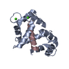
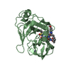
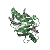
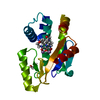
- PDB-4pww: Crystal Structure of Engineered Protein. Northeast Structural Gen... -
+ Open data

- Basic information
| Entry | Database: PDB / ID: 4pww | ||||||
|---|---|---|---|---|---|---|---|
| Title | Crystal Structure of Engineered Protein. Northeast Structural Genomics Consortium Target OR494. | ||||||
 Components Components | OR494 | ||||||
 Keywords Keywords | DE NOVO PROTEIN / Structural Genomics / PSI-Biology / Protein Structure Initiative / Northeast Structural Genomics Consortium / NESG / De Novo / OR494 | ||||||
| Function / homology | Ribosomal protein S10 / Alpha-Beta Plaits / 2-Layer Sandwich / Alpha Beta / ACETIC ACID / PHOSPHATE ION Function and homology information Function and homology information | ||||||
| Biological species | synthetic construct (others) | ||||||
| Method |  X-RAY DIFFRACTION / SYNCHROTRON / MOLECULAR REPLACEMENT / Resolution: 1.471 Å X-RAY DIFFRACTION / SYNCHROTRON / MOLECULAR REPLACEMENT / Resolution: 1.471 Å | ||||||
 Authors Authors | Vorobiev, S. / Lin, Y.-R. / Seetharaman, J. / Xiao, R. / Everett, J.K. / Acton, T.B. / Baker, D. / Montelione, G.T. / Tong, L. / Hunt, J.F. / Northeast Structural Genomics Consortium (NESG) | ||||||
 Citation Citation | Journal: To be Published Title: Crystal Structure of Engineered Protein OR494. Authors: Vorobiev, S. / Lin, Y.-R. / Seetharaman, J. / Xiao, R. / Everett, J.K. / Acton, T.B. / Baker, D. / Montelione, G.T. / Tong, L. / Hunt, J.F. | ||||||
| History |
|
- Structure visualization
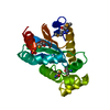
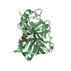
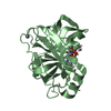
| Structure viewer | Molecule: MolmilJmol/JSmol |
|---|
- Downloads & links
-Download
| PDBx/mmCIF format | 4pww.cif.gz | 49.3 KB | Display | PDBx/mmCIF format |
|---|---|---|---|---|
| PDB format | pdb4pww.ent.gz | 34.9 KB | Display | PDB format |
| PDBx/mmJSON format | 4pww.json.gz | Tree view | PDBx/mmJSON format | |
| Others |  Other downloads Other downloads |
-Validation report
| Arichive directory | https://data.pdbj.org/pub/pdb/validation_reports/pw/4pwwftp://data.pdbj.org/pub/pdb/validation_reports/pw/4pww | HTTPS FTP |
|---|
-Related structure data
| Related structure data |  4ky3S S: Starting model for refinement |
|---|---|
| Similar structure data | |
| Other databases |


-Links
 PDBj
PDBj


- Assembly
| Deposited unit | 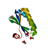
| ||||||||||||
|---|---|---|---|---|---|---|---|---|---|---|---|---|---|
| 1 | 
| ||||||||||||
| Unit cell |
| ||||||||||||
| Components on special symmetry positions |
| ||||||||||||
| Details | dimer as reported by HPLC+light scattering |
-Components
| #1: Protein | Mass: 10190.724 Da / Num. of mol.: 1 Source method: isolated from a genetically manipulated source Source: (gene. exp.) synthetic construct (others) / Plasmid: OR494-21.1 / Production host:  | ||
|---|---|---|---|
| #2: Chemical | ChemComp-PO4 /   Mass: 94.971 Da / Num. of mol.: 1 / Source method: obtained synthetically / Formula: PO4 Mass: 94.971 Da / Num. of mol.: 1 / Source method: obtained synthetically / Formula: PO4 | ||
| #3: Chemical |   Mass: 60.052 Da / Num. of mol.: 3 / Source method: obtained synthetically / Formula: C2H4O2 Mass: 60.052 Da / Num. of mol.: 3 / Source method: obtained synthetically / Formula: C2H4O2#4: Water | ChemComp-HOH / |  Mass: 18.015 Da / Num. of mol.: 96 / Source method: isolated from a natural source / Formula: H2O Mass: 18.015 Da / Num. of mol.: 96 / Source method: isolated from a natural source / Formula: H2O |
-Experimental details
-Experiment
| Experiment | Method: X-RAY DIFFRACTION / Number of used crystals: 1 |
|---|
- Sample preparation
| Crystal | Density Matthews: 2.1 Å3/Da / Density % sol: 41.32 % |
|---|---|
| Crystal grow | Temperature: 291 K / Method: microbatch crystallization under oil / pH: 3.5 Details: Protein solution: 100mM NaCl, 5mM DTT, 0.02% NaN3, 10mM Tris-HCl (pH 7.5). Reservoir solution: 2.0M ammonium sulfate, 0.1M citric acid, pH 3.5, Microbatch crystallization under oil, temperature 291K |
-Data collection
| Diffraction | Mean temperature: 100 K |
|---|---|
| Diffraction source | Source: SYNCHROTRON / Site: NSLS  / Beamline: X4C / Wavelength: 0.97907 Å / Beamline: X4C / Wavelength: 0.97907 Å |
| Detector | Type: MAR CCD 165 mm / Detector: CCD / Date: Mar 14, 2014 |
| Radiation | Monochromator: Si 111 CHANNEL / Protocol: SINGLE WAVELENGTH / Monochromatic (M) / Laue (L): M / Scattering type: x-ray |
| Radiation wavelength | Wavelength: 0.97907 Å / Relative weight: 1 |
| Reflection | Resolution: 1.47→50 Å / Num. all: 28181 / Num. obs: 27476 / % possible obs: 97.5 % / Observed criterion σ(F): 0 / Observed criterion σ(I): 0 / Redundancy: 4.5 % / Biso Wilson estimate: 16.38 Å2 / Rmerge(I) obs: 0.055 / Net I/σ(I): 33.25 |
| Reflection shell | Resolution: 1.47→1.52 Å / Redundancy: 3.7 % / Rmerge(I) obs: 0.403 / Mean I/σ(I) obs: 4.2 / Num. unique all: 2840 / % possible all: 91.2 |
- Processing
| Software |
| |||||||||||||||||||||||||||||||||||||||||||||||||||||||||||||||||||||||||||||
|---|---|---|---|---|---|---|---|---|---|---|---|---|---|---|---|---|---|---|---|---|---|---|---|---|---|---|---|---|---|---|---|---|---|---|---|---|---|---|---|---|---|---|---|---|---|---|---|---|---|---|---|---|---|---|---|---|---|---|---|---|---|---|---|---|---|---|---|---|---|---|---|---|---|---|---|---|---|---|
| Refinement | Method to determine structure: MOLECULAR REPLACEMENT Starting model: PDB ENTRY 4KY3 Resolution: 1.471→36.569 Å / Occupancy max: 1 / Occupancy min: 0.43 / SU ML: 0.32 / Cross valid method: THROUGHOUT / σ(F): 1.91 / Phase error: 22.04 / Stereochemistry target values: ML Details: Presence of phosphate and acetate ions were traced back to a purification procedure. There is unknown ligand in the structure which was modeled by water molecules (A593-596).
| |||||||||||||||||||||||||||||||||||||||||||||||||||||||||||||||||||||||||||||
| Solvent computation | Shrinkage radii: 0.9 Å / VDW probe radii: 1.11 Å / Solvent model: FLAT BULK SOLVENT MODEL / Bsol: 50.118 Å2 / ksol: 0.379 e/Å3 | |||||||||||||||||||||||||||||||||||||||||||||||||||||||||||||||||||||||||||||
| Displacement parameters | Biso max: 64.69 Å2 / Biso mean: 25.281 Å2 / Biso min: 11 Å2
| |||||||||||||||||||||||||||||||||||||||||||||||||||||||||||||||||||||||||||||
| Refinement step | Cycle: LAST / Resolution: 1.471→36.569 Å
| |||||||||||||||||||||||||||||||||||||||||||||||||||||||||||||||||||||||||||||
| Refine LS restraints |
| |||||||||||||||||||||||||||||||||||||||||||||||||||||||||||||||||||||||||||||
| LS refinement shell | Refine-ID: X-RAY DIFFRACTION / Total num. of bins used: 10
|
