 Movie
Movie Controller


+ Open data

- Basic information
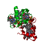
| Entry | Database: PDB / ID: 1cm4 | ||||||
|---|---|---|---|---|---|---|---|
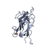
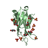
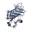
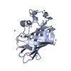
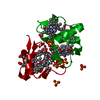
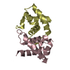
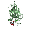
| Title | Motions of calmodulin-four-conformer refinement | ||||||
 Components Components |
| ||||||
 Keywords Keywords | CALCIUM-BINDING/TRANSFERASE / EF-HAND CALCIUM-BINDING PROTEIN / CALCIUM-BINDING-TRANSFERASE complex | ||||||
| Function / homology |  Function and homology information Function and homology information: / HSF1-dependent transactivation / RAF activation / regulation of store-operated calcium channel activity / : / Ion transport by P-type ATPases / peptidyl-threonine autophosphorylation / : / calcium- and calmodulin-dependent protein kinase complex / regulation of endocannabinoid signaling pathway ...: / HSF1-dependent transactivation / RAF activation / regulation of store-operated calcium channel activity / : / Ion transport by P-type ATPases / peptidyl-threonine autophosphorylation / : / calcium- and calmodulin-dependent protein kinase complex / regulation of endocannabinoid signaling pathway / neurotransmitter receptor transport to plasma membrane / Interferon gamma signaling / regulation of response to tumor cell / positive regulation of autophagic cell death / DAPK1-calmodulin complex / Ca2+/calmodulin-dependent protein kinase / : / : / : / : / dendritic spine development / : / negative regulation of hydrolase activity / regulation of neurotransmitter secretion / Trafficking of AMPA receptors / : / type 3 metabotropic glutamate receptor binding / positive regulation of calcium ion transport / regulation of neuron migration / establishment of protein localization to membrane / calcium/calmodulin-dependent protein kinase activity / Ca2+ pathway / regulation of mitochondrial membrane permeability involved in apoptotic process / RAF/MAP kinase cascade / GTPase activating protein binding / positive regulation of DNA binding / Ion homeostasis / dendrite morphogenesis / negative regulation of high voltage-gated calcium channel activity / negative regulation of ryanodine-sensitive calcium-release channel activity / organelle localization by membrane tethering / : / autophagosome membrane docking / regulation of synaptic vesicle exocytosis / negative regulation of calcium ion export across plasma membrane / regulation of ryanodine-sensitive calcium-release channel activity / regulation of cardiac muscle cell action potential / regulation of neurotransmitter receptor localization to postsynaptic specialization membrane / negative regulation of ferroptosis / calcineurin-mediated signaling / nitric-oxide synthase binding / adenylate cyclase binding / protein phosphatase activator activity / Unblocking of NMDA receptors, glutamate binding and activation / regulation of synaptic vesicle endocytosis / regulation of neuronal synaptic plasticity / glutamate receptor binding / detection of calcium ion / postsynaptic cytosol / regulation of cardiac muscle contraction / cell surface receptor signaling pathway via JAK-STAT / catalytic complex / regulation of protein localization to plasma membrane / positive regulation of nitric-oxide synthase activity / phosphatidylinositol 3-kinase binding / activation of adenylate cyclase activity / calcium channel inhibitor activity / presynaptic cytosol / cellular response to interferon-beta / regulation of release of sequestered calcium ion into cytosol by sarcoplasmic reticulum / enzyme regulator activity / titin binding / regulation of calcium-mediated signaling / voltage-gated potassium channel complex / positive regulation of cardiac muscle cell apoptotic process / calcium channel complex / potassium ion transmembrane transport / ionotropic glutamate receptor signaling pathway / regulation of heart rate / dendrite cytoplasm / nitric-oxide synthase regulator activity / response to ischemia / adenylate cyclase activator activity / angiotensin-activated signaling pathway / regulation of cytokinesis / spindle microtubule / positive regulation of receptor signaling pathway via JAK-STAT / response to amphetamine / sarcomere / calcium channel regulator activity / calcium-mediated signaling / G1/S transition of mitotic cell cycle / response to calcium ion / cellular response to type II interferon / G2/M transition of mitotic cell cycle / Schaffer collateral - CA1 synapse / spindle pole / disordered domain specific binding / calcium-dependent protein binding / kinase activity Similarity search - Function | ||||||
| Biological species |  | ||||||
| Method |  X-RAY DIFFRACTION / SYNCHROTRON / MOLECULAR REPLACEMENT / Resolution: 2 Å X-RAY DIFFRACTION / SYNCHROTRON / MOLECULAR REPLACEMENT / Resolution: 2 Å | ||||||
 Authors Authors | Wall, M.E. / Phillips Jr., G.N. | ||||||
 Citation Citation | Journal: Structure / Year: 1997 Title: Motions of calmodulin characterized using both Bragg and diffuse X-ray scattering. Authors: Wall, M.E. / Clarage, J.B. / Phillips Jr., G.N. #1: Journal: Science / Year: 1993Title: Modulation of Calmodulin Plasticity in Molecular Recognition on the Basis of X-Ray Structures Authors: Meador, W.E. / Means, A.R. / Quiocho, F.A. #2: Journal: Science / Year: 1992Title: Target Enzyme Recognition by Calmodulin: 2.4 A Structure of a Calmodulin-Peptide Complex Authors: Meador, W.E. / Means, A.R. / Quiocho, F.A. | ||||||
| History |
|
- Structure visualization
| Structure viewer | Molecule: MolmilJmol/JSmol |
|---|
- Downloads & links
-Download
| PDBx/mmCIF format | 1cm4.cif.gz | 119.8 KB | Display | PDBx/mmCIF format |
|---|---|---|---|---|
| PDB format | pdb1cm4.ent.gz | 102.4 KB | Display | PDB format |
| PDBx/mmJSON format | 1cm4.json.gz | Tree view | PDBx/mmJSON format | |
| Others |  Other downloads Other downloads |
-Validation report
| Arichive directory | https://data.pdbj.org/pub/pdb/validation_reports/cm/1cm4ftp://data.pdbj.org/pub/pdb/validation_reports/cm/1cm4 | HTTPS FTP |
|---|
-Related structure data
| Related structure data | 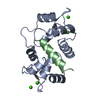 1cm1SC S: Starting model for refinement C: citing same article ( |
|---|---|
| Similar structure data |


-Links
 PDBj
PDBj


- Assembly
| Deposited unit | 
| ||||||||
|---|---|---|---|---|---|---|---|---|---|
| 1 |
| ||||||||
| Unit cell |
| ||||||||
| Components on special symmetry positions |
|
-Components
| #1: Protein | Mass: 16721.350 Da / Num. of mol.: 1 / Source method: isolated from a natural source / Details: SIGMA LOT 54H9558 / Source: (natural) | ||
|---|---|---|---|
| #2: Protein/peptide | Mass: 2886.528 Da / Num. of mol.: 1 / Fragment: CALMODULIN BINDING DOMAIN, RESIDUES 290 - 314 / Source method: obtained synthetically / References: UniProt: P11275, EC: 2.7.1.123 | ||
| #3: Chemical | ChemComp-CA /   Mass: 40.078 Da / Num. of mol.: 17 / Source method: obtained synthetically / Formula: Ca Mass: 40.078 Da / Num. of mol.: 17 / Source method: obtained synthetically / Formula: Ca#4: Water | ChemComp-HOH / |  Mass: 18.015 Da / Num. of mol.: 58 / Source method: isolated from a natural source / Formula: H2O Mass: 18.015 Da / Num. of mol.: 58 / Source method: isolated from a natural source / Formula: H2O |
-Experimental details
-Experiment
| Experiment | Method: X-RAY DIFFRACTION / Number of used crystals: 1 |
|---|
- Sample preparation
| Crystal | Density Matthews: 2.23 Å3/Da / Density % sol: 44.86 % | ||||||||||||||||||||||||||||||||||||||||||||||||
|---|---|---|---|---|---|---|---|---|---|---|---|---|---|---|---|---|---|---|---|---|---|---|---|---|---|---|---|---|---|---|---|---|---|---|---|---|---|---|---|---|---|---|---|---|---|---|---|---|---|
| Crystal grow | Method: vapor diffusion - hanging drop - microseeding / pH: 5.2 Details: DIFFRACTION-QUALITY CRYSTALS WERE MICROSEEDED IN HANGING DROPS OVER 100 MM SODIUM ACETATE AT PH 5.2, WITH 20% POLY-ETHYLENE GLYCOL 6000 (PEG 6000), 10 MM CALCIUM CHLORIDE AND 0.02% SODIUM ...Details: DIFFRACTION-QUALITY CRYSTALS WERE MICROSEEDED IN HANGING DROPS OVER 100 MM SODIUM ACETATE AT PH 5.2, WITH 20% POLY-ETHYLENE GLYCOL 6000 (PEG 6000), 10 MM CALCIUM CHLORIDE AND 0.02% SODIUM AZIDE. STOCK SOLUTIONS OF 24 MG/ML BOVINE BRAIN CALMODULIN (SIGMA LOT 54H9558), 14 MG/ML CAMKII-ALPHA PEPTIDE, AND 30% PEG WERE MIXED INTO HANGING DROPS IN ABOUT A 4-2-1 RATIO., vapor diffusion - hanging drop - microseeding | ||||||||||||||||||||||||||||||||||||||||||||||||
| Crystal grow | *PLUS Method: vapor diffusion, hanging drop / Details: used to seeding | ||||||||||||||||||||||||||||||||||||||||||||||||
| Components of the solutions | *PLUS
|
-Data collection
| Diffraction | Mean temperature: 302 K |
|---|---|
| Diffraction source | Source: SYNCHROTRON / Site: CHESS  / Beamline: F2 / Wavelength: 0.98 / Beamline: F2 / Wavelength: 0.98 |
| Detector | Type: PRINCETON 2K / Detector: CCD / Date: May 1, 1996 / Details: MIRRORS |
| Radiation | Monochromator: SINGLE-CRYSTAL / Monochromatic (M) / Laue (L): M / Scattering type: x-ray |
| Radiation wavelength | Wavelength: 0.98 Å / Relative weight: 1 |
| Reflection | Resolution: 2→10 Å / Num. obs: 11522 / % possible obs: 92.1 % / Observed criterion σ(I): 2 / Biso Wilson estimate: 22.5 Å2 / Rsym value: 0.061 / Net I/σ(I): 9.2 |
| Reflection shell | Resolution: 2→2.07 Å / Rsym value: 0.24 / % possible all: 82.8 |
- Processing
| Software |
| ||||||||||||||||||||||||||||||||||||||||||||||||||||||||||||||||||||||||||||||||
|---|---|---|---|---|---|---|---|---|---|---|---|---|---|---|---|---|---|---|---|---|---|---|---|---|---|---|---|---|---|---|---|---|---|---|---|---|---|---|---|---|---|---|---|---|---|---|---|---|---|---|---|---|---|---|---|---|---|---|---|---|---|---|---|---|---|---|---|---|---|---|---|---|---|---|---|---|---|---|---|---|---|
| Refinement | Method to determine structure: MOLECULAR REPLACEMENT Starting model: PDB ENTRY 1CM1 Resolution: 2→10 Å / Rfactor Rfree error: 0.008 / Data cutoff high absF: 100000 / Data cutoff low absF: 0.1 / Isotropic thermal model: RESTRAINED / Cross valid method: THROUGHOUT / σ(F): 2 Details: RESIDUES 74 - 83, WHICH ARE NOT PRESENT IN PDB ENTRY 1CDM, WERE ADDED FOR THIS REFINEMENT. AN INITIAL GUESS WAS OBTAINED FROM THE COORDINATES OF PDB ENTRY 1CDL. THESE RESIDUES DO NOT SHOW ...Details: RESIDUES 74 - 83, WHICH ARE NOT PRESENT IN PDB ENTRY 1CDM, WERE ADDED FOR THIS REFINEMENT. AN INITIAL GUESS WAS OBTAINED FROM THE COORDINATES OF PDB ENTRY 1CDL. THESE RESIDUES DO NOT SHOW CONNECTED ELECTRON DENSITY AT A LEVEL OF 1SIGMA.
| ||||||||||||||||||||||||||||||||||||||||||||||||||||||||||||||||||||||||||||||||
| Displacement parameters | Biso mean: 29.1 Å2 | ||||||||||||||||||||||||||||||||||||||||||||||||||||||||||||||||||||||||||||||||
| Refine analyze |
| ||||||||||||||||||||||||||||||||||||||||||||||||||||||||||||||||||||||||||||||||
| Refinement step | Cycle: LAST / Resolution: 2→10 Å
| ||||||||||||||||||||||||||||||||||||||||||||||||||||||||||||||||||||||||||||||||
| Refine LS restraints |
| ||||||||||||||||||||||||||||||||||||||||||||||||||||||||||||||||||||||||||||||||
| LS refinement shell | Resolution: 2→2.07 Å / Rfactor Rfree error: 0.023 / Total num. of bins used: 10
| ||||||||||||||||||||||||||||||||||||||||||||||||||||||||||||||||||||||||||||||||
| Xplor file |
| ||||||||||||||||||||||||||||||||||||||||||||||||||||||||||||||||||||||||||||||||
| Software | *PLUS Name: X-PLOR / Version: 3.851 / Classification: refinement | ||||||||||||||||||||||||||||||||||||||||||||||||||||||||||||||||||||||||||||||||
| Refine LS restraints | *PLUS
|
