 ムービー
ムービー コントローラー


+ データを開く

- 基本情報
基本情報
| 登録情報 | データベース: PDB / ID: 1qe1 | ||||||
|---|---|---|---|---|---|---|---|
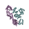
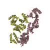
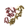
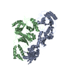
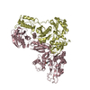
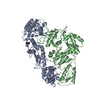
| タイトル | CRYSTAL STRUCTURE OF 3TC-RESISTANT M184I MUTANT OF HIV-1 REVERSE TRANSCRIPTASE | ||||||
 要素 要素 |
| ||||||
 キーワード キーワード | TRANSFERASE / HIV / REVERSE TRANSCRIPTASE / 3TC / RESISTANCE / MUTANT / DNA POLYMERASE | ||||||
| 機能・相同性 |  機能・相同性情報 機能・相同性情報HIV-1 retropepsin / symbiont-mediated activation of host apoptosis / retroviral ribonuclease H / exoribonuclease H / exoribonuclease H activity / host multivesicular body / DNA integration / viral genome integration into host DNA / RNA-directed DNA polymerase / establishment of integrated proviral latency ...HIV-1 retropepsin / symbiont-mediated activation of host apoptosis / retroviral ribonuclease H / exoribonuclease H / exoribonuclease H activity / host multivesicular body / DNA integration / viral genome integration into host DNA / RNA-directed DNA polymerase / establishment of integrated proviral latency / viral penetration into host nucleus / RNA stem-loop binding / RNA-directed DNA polymerase activity / RNA-DNA hybrid ribonuclease activity / 転移酵素; リンを含む基を移すもの; 核酸を移すもの / host cell / viral nucleocapsid / DNA recombination / DNA-directed DNA polymerase / aspartic-type endopeptidase activity / 加水分解酵素; エステル加水分解酵素 / DNA-directed DNA polymerase activity / symbiont-mediated suppression of host gene expression / viral translational frameshifting / lipid binding / symbiont entry into host cell / host cell nucleus / host cell plasma membrane / virion membrane / structural molecule activity / proteolysis / DNA binding / zinc ion binding / membrane 類似検索 - 分子機能 | ||||||
| 生物種 |  Human immunodeficiency virus type 1 BH10 (ヒト免疫不全ウイルス) Human immunodeficiency virus type 1 BH10 (ヒト免疫不全ウイルス) | ||||||
| 手法 |  X線回折 / シンクロトロン / 解像度: 2.85 Å X線回折 / シンクロトロン / 解像度: 2.85 Å | ||||||
 データ登録者 データ登録者 | Sarafianos, S.G. / Das, K. / Ding, J. / Hughes, S.H. / Arnold, E. | ||||||
 引用 引用 | ジャーナル: Proc.Natl.Acad.Sci.USA / 年: 1999 タイトル: Lamivudine (3TC) resistance in HIV-1 reverse transcriptase involves steric hindrance with beta-branched amino acids. 著者: Sarafianos, S.G. / Das, K. / Clark Jr., A.D. / Ding, J. / Boyer, P.L. / Hughes, S.H. / Arnold, E. #1: ジャーナル: Structure / 年: 1996タイトル: Structure of Unliganded HIV-1 Reverse Transcriptase at 2.7 A resolution: Implications of conformational changes for polymerization and inhibition mechanisms 著者: Hsiou, Y. / Ding, J. / Das, K. / Clark Jr., A.D. / Hughes, S.H. / Arnold, E. #2: ジャーナル: J.Mol.Biol. / 年: 1998タイトル: Structure and Functional Implications of the Polymerase Active Site Region in a Complex of HIV-1 RT with a Double-Stranded DNA and an Antibody Fab Fragment at 2.8 Angstroms Resolution 著者: Ding, J. / Das, K. / Hsiou, Y. / Sarafianos, S.G. / Clark Jr., A.D. / Jacobo-Molina, A. / Tantillo, C. / Hughes, S.H. / Arnold, E. | ||||||
| 履歴 |
|
- 構造の表示
構造の表示
| 構造ビューア | 分子: MolmilJmol/JSmol |
|---|
- ダウンロードとリンク
-ダウンロード
| PDBx/mmCIF形式 | 1qe1.cif.gz | 199.7 KB | 表示 | PDBx/mmCIF形式 |
|---|---|---|---|---|
| PDB形式 | pdb1qe1.ent.gz | 160.7 KB | 表示 | PDB形式 |
| PDBx/mmJSON形式 | 1qe1.json.gz | ツリー表示 | PDBx/mmJSON形式 | |
| その他 |  その他のダウンロード その他のダウンロード |
-検証レポート
| 文書・要旨 | 1qe1_validation.pdf.gz | 441.5 KB | 表示 | wwPDB検証レポート |
|---|---|---|---|---|
| 文書・詳細版 | 1qe1_full_validation.pdf.gz | 493.7 KB | 表示 | |
| XML形式データ | 1qe1_validation.xml.gz | 40.3 KB | 表示 | |
| CIF形式データ | 1qe1_validation.cif.gz | 54 KB | 表示 | |
| アーカイブディレクトリ | https://data.pdbj.org/pub/pdb/validation_reports/qe/1qe1ftp://data.pdbj.org/pub/pdb/validation_reports/qe/1qe1 | HTTPS FTP |
-関連構造データ


-リンク
 PDBj
PDBj


- 集合体
集合体
| 登録構造単位 | 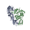
| ||||||||||
|---|---|---|---|---|---|---|---|---|---|---|---|
| 1 |
| ||||||||||
| 単位格子 |
| ||||||||||
| 詳細 | Heterodimer, of two subunits, P66 and P51. P51 contains the N-terminal 440 residues of the P66 subunit. |
-要素
| #1: タンパク質 | 分子量: 64256.613 Da / 分子数: 1 / 断片: SUBUNIT A (P66), RESIDUES 168-725 / 変異: M184I,C280S / 由来タイプ: 組換発現 由来: (組換発現) Human immunodeficiency virus type 1 BH10 (ヒト免疫不全ウイルス)属: Lentivirus / 生物種: Human immunodeficiency virus 1 / 発現宿主:  |
|---|---|
| #2: タンパク質 | 分子量: 49893.320 Da / 分子数: 1 / 断片: SUBUNIT B (P51), RESIDUES 168-594 / 変異: M184I,C280S / 由来タイプ: 組換発現 由来: (組換発現) Human immunodeficiency virus type 1 BH10 (ヒト免疫不全ウイルス)属: Lentivirus / 生物種: Human immunodeficiency virus 1 / 発現宿主: |
-実験情報
-実験
| 実験 | 手法: X線回折 / 使用した結晶の数: 1 |
|---|
- 試料調製
試料調製
| 結晶 | マシュー密度: 3.48 Å3/Da / 溶媒含有率: 64.7 % | |||||||||||||||||||||||||
|---|---|---|---|---|---|---|---|---|---|---|---|---|---|---|---|---|---|---|---|---|---|---|---|---|---|---|
| 結晶化 | 温度: 277 K / 手法: 蒸気拡散法, ハンギングドロップ法 / pH: 6.8 詳細: PEG 8000, GLYCEROL, BIS-TRIS, AMMONIUM SULFATE, pH 6.8, VAPOR DIFFUSION, HANGING DROP, temperature 277K | |||||||||||||||||||||||||
| 結晶化 | *PLUS | |||||||||||||||||||||||||
| 溶液の組成 | *PLUS
|
-データ収集
| 回折 | 平均測定温度: 108 K |
|---|---|
| 放射光源 | 由来: シンクロトロン / サイト: CHESS  / ビームライン: F1 / 波長: 0.928 / ビームライン: F1 / 波長: 0.928 |
| 検出器 | タイプ: PRINCETON 2K / 検出器: CCD |
| 放射 | プロトコル: SINGLE WAVELENGTH / 単色(M)・ラウエ(L): M / 散乱光タイプ: x-ray |
| 放射波長 | 波長: 0.928 Å / 相対比: 1 |
| 反射 | 解像度: 2.85→40 Å / Num. all: 37000 / Num. obs: 35177 / % possible obs: 95 % / Observed criterion σ(I): 0 / Rmerge(I) obs: 0.09 / Net I/σ(I): 5.2 |
| 反射 シェル | 解像度: 2.85→2.95 Å / 冗長度: 2.5 % / Rmerge(I) obs: 0.36 / % possible all: 88 |
| 反射 | *PLUS 最低解像度: 40 Å / % possible obs: 95 % / Rmerge(I) obs: 0.09 |
| 反射 シェル | *PLUS % possible obs: 88 % |
- 解析
解析
| ソフトウェア |
| ||||||||||||||||||||
|---|---|---|---|---|---|---|---|---|---|---|---|---|---|---|---|---|---|---|---|---|---|
| 精密化 | 解像度: 2.85→8 Å / 交差検証法: THROUGHOUT / σ(I): 1 / 立体化学のターゲット値: Engh & Huber
| ||||||||||||||||||||
| 精密化ステップ | サイクル: LAST / 解像度: 2.85→8 Å
| ||||||||||||||||||||
| 拘束条件 |
| ||||||||||||||||||||
| ソフトウェア | *PLUS 名称: X-PLOR / バージョン: 3.843 / 分類: refinement | ||||||||||||||||||||
| 精密化 | *PLUS 最低解像度: 8 Å / Rfactor obs: 0.259 | ||||||||||||||||||||
| 溶媒の処理 | *PLUS | ||||||||||||||||||||
| 原子変位パラメータ | *PLUS |
