 Movie
Movie Controller

[English] 日本語
 Yorodumi
- PDB-1q9a: Crystal structure of the sarcin/ricin domain from E.coli 23S rRNA... -
+ Open data

- Basic information
| Entry | Database: PDB / ID: 1q9a | ||||||
|---|---|---|---|---|---|---|---|
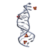
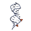
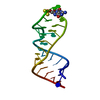
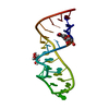
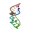
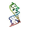
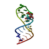
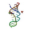
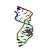
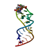
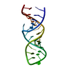
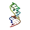
| Title | Crystal structure of the sarcin/ricin domain from E.coli 23S rRNA at 1.04 resolution | ||||||
 Components Components | Sarcin/ricin 23S rRNA | ||||||
 Keywords Keywords | RNA / sarcin/ricin domain / ribonucleic acid / RNA recognition / high resolution / ribosomes | ||||||
| Function / homology | : / RNA / RNA (> 10) Function and homology information Function and homology information | ||||||
| Method |  X-RAY DIFFRACTION / SYNCHROTRON / FOURIER SYNTHESIS / Resolution: 1.04 Å X-RAY DIFFRACTION / SYNCHROTRON / FOURIER SYNTHESIS / Resolution: 1.04 Å | ||||||
 Authors Authors | Correll, C.C. / Beneken, J. / Plantinga, M.J. / Lubbers, M. / Chan, Y.L. | ||||||
 Citation Citation | Journal: Nucleic Acids Res. / Year: 2003 Title: The common and distinctive features of the bulged-G motif based on a 1.04 A resolution RNA structure Authors: Correll, C.C. / Beneken, J. / Plantinga, M.J. / Lubbers, M. / Chan, Y.L. | ||||||
| History |
|
- Structure visualization
| Structure viewer | Molecule: MolmilJmol/JSmol |
|---|
- Downloads & links
-Download
| PDBx/mmCIF format | 1q9a.cif.gz | 49.5 KB | Display | PDBx/mmCIF format |
|---|---|---|---|---|
| PDB format | pdb1q9a.ent.gz | 37.7 KB | Display | PDB format |
| PDBx/mmJSON format | 1q9a.json.gz | Tree view | PDBx/mmJSON format | |
| Others |  Other downloads Other downloads |
-Validation report
| Arichive directory | https://data.pdbj.org/pub/pdb/validation_reports/q9/1q9aftp://data.pdbj.org/pub/pdb/validation_reports/q9/1q9a | HTTPS FTP |
|---|
-Related structure data


-Links
 PDBj
PDBj


- Assembly
| Deposited unit | 
| ||||||||
|---|---|---|---|---|---|---|---|---|---|
| 1 |
| ||||||||
| Unit cell |
|
-Components
| #1: RNA chain | Mass: 8744.255 Da / Num. of mol.: 1 / Fragment: sarcin/ricin domain / Source method: obtained synthetically Details: THE 23S ribosomal RNA WAS CHEMICALLY SYNTHESIZED. THE SEQUENCE OF THE 23S ribosomal RNA IS NATURALLY FOUND IN Escherichia coli (bacteria). References: GenBank: 26111017 |
|---|---|
| #2: Water | ChemComp-HOH /  Mass: 18.015 Da / Num. of mol.: 179 / Source method: isolated from a natural source / Formula: H2O Mass: 18.015 Da / Num. of mol.: 179 / Source method: isolated from a natural source / Formula: H2O |
-Experimental details
-Experiment
| Experiment | Method: X-RAY DIFFRACTION / Number of used crystals: 1 |
|---|
- Sample preparation
| Crystal | Density Matthews: 1.91 Å3/Da / Density % sol: 35.57 % | |||||||||||||||||||||||||||||||||||||||||||||||||||||||||||||||||||||||||||||
|---|---|---|---|---|---|---|---|---|---|---|---|---|---|---|---|---|---|---|---|---|---|---|---|---|---|---|---|---|---|---|---|---|---|---|---|---|---|---|---|---|---|---|---|---|---|---|---|---|---|---|---|---|---|---|---|---|---|---|---|---|---|---|---|---|---|---|---|---|---|---|---|---|---|---|---|---|---|---|
| Crystal grow | Temperature: 295 K / Method: vapor diffusion, hanging drop / pH: 7 Details: ammonium sulfate, K-MOPS, magnesium chloride, manganese chloride, pH 7.0, VAPOR DIFFUSION, HANGING DROP, temperature 295K | |||||||||||||||||||||||||||||||||||||||||||||||||||||||||||||||||||||||||||||
| Components of the solutions |
| |||||||||||||||||||||||||||||||||||||||||||||||||||||||||||||||||||||||||||||
| Crystal grow | *PLUS Method: vapor diffusion | |||||||||||||||||||||||||||||||||||||||||||||||||||||||||||||||||||||||||||||
| Components of the solutions | *PLUS
|
-Data collection
| Diffraction | Mean temperature: 100 K |
|---|---|
| Diffraction source | Source: SYNCHROTRON / Site: APS  / Beamline: 19-ID / Wavelength: 0.97764 Å / Beamline: 19-ID / Wavelength: 0.97764 Å |
| Detector | Type: CUSTOM-MADE / Detector: CCD / Date: Jul 7, 2000 |
| Radiation | Protocol: SINGLE WAVELENGTH / Monochromatic (M) / Laue (L): M / Scattering type: x-ray |
| Radiation wavelength | Wavelength: 0.97764 Å / Relative weight: 1 |
| Reflection | Resolution: 1.04→40 Å / Num. all: 31452 / Num. obs: 29848 / % possible obs: 94.9 % / Observed criterion σ(F): 0 / Observed criterion σ(I): 0 / Redundancy: 5.1 % / Rmerge(I) obs: 0.037 / Net I/σ(I): 55.3 |
| Reflection shell | Resolution: 1.04→1.06 Å / Rmerge(I) obs: 0.446 / Mean I/σ(I) obs: 2 / % possible all: 82.5 |
| Reflection shell | *PLUS % possible obs: 82.5 % |
- Processing
| Software |
| ||||||||||||||||||||||||||||||||||||||||||||||||||||||||||||
|---|---|---|---|---|---|---|---|---|---|---|---|---|---|---|---|---|---|---|---|---|---|---|---|---|---|---|---|---|---|---|---|---|---|---|---|---|---|---|---|---|---|---|---|---|---|---|---|---|---|---|---|---|---|---|---|---|---|---|---|---|---|
| Refinement | Method to determine structure: FOURIER SYNTHESIS / Resolution: 1.04→40 Å / σ(F): 0 / σ(I): 0 / Stereochemistry target values: Engh & Huber
| ||||||||||||||||||||||||||||||||||||||||||||||||||||||||||||
| Refinement step | Cycle: LAST / Resolution: 1.04→40 Å
| ||||||||||||||||||||||||||||||||||||||||||||||||||||||||||||
| Refine LS restraints |
| ||||||||||||||||||||||||||||||||||||||||||||||||||||||||||||
| Software | *PLUS Name: SHELXL / Version: 97 / Classification: refinement | ||||||||||||||||||||||||||||||||||||||||||||||||||||||||||||
| Refine LS restraints | *PLUS
| ||||||||||||||||||||||||||||||||||||||||||||||||||||||||||||
| LS refinement shell | *PLUS Rfactor Rfree: 0.319 |
