 Movie
Movie Controller

[English] 日本語
 Yorodumi
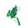
- PDB-1jbr: Crystal Structure of the Ribotoxin Restrictocin and a 31-mer SRD ... -
+ Open data

- Basic information
| Entry | Database: PDB / ID: 1jbr | ||||||
|---|---|---|---|---|---|---|---|
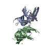
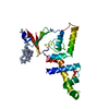
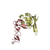
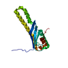
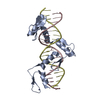
| Title | Crystal Structure of the Ribotoxin Restrictocin and a 31-mer SRD RNA Inhibitor | ||||||
 Components Components |
| ||||||
 Keywords Keywords | HYDROLASE/RNA / protein-RNA interaction / specific recognition / restrictocin / ribosomal RNA / sarcin/ricin domain / base flipping / HYDROLASE-RNA COMPLEX | ||||||
| Function / homology |  Function and homology information Function and homology informationHydrolases; Acting on ester bonds; Endoribonucleases producing 3'-phosphomonoesters / RNA endonuclease activity / negative regulation of translation / hydrolase activity / RNA binding / extracellular region Similarity search - Function | ||||||
| Biological species |  | ||||||
| Method |  X-RAY DIFFRACTION / SYNCHROTRON / MOLECULAR REPLACEMENT / Resolution: 2.15 Å X-RAY DIFFRACTION / SYNCHROTRON / MOLECULAR REPLACEMENT / Resolution: 2.15 Å | ||||||
 Authors Authors | Yang, X. / Gerczei, T. / Glover, L. / Correll, C.C. | ||||||
 Citation Citation | Journal: Nat.Struct.Biol. / Year: 2001 Title: Crystal structures of restrictocin-inhibitor complexes with implications for RNA recognition and base flipping. Authors: Yang, X. / Gerczei, T. / Glover, L.T. / Correll, C.C. | ||||||
| History |
|
- Structure visualization
| Structure viewer | Molecule: MolmilJmol/JSmol |
|---|
- Downloads & links
-Download
| PDBx/mmCIF format | 1jbr.cif.gz | 112.8 KB | Display | PDBx/mmCIF format |
|---|---|---|---|---|
| PDB format | pdb1jbr.ent.gz | 82.9 KB | Display | PDB format |
| PDBx/mmJSON format | 1jbr.json.gz | Tree view | PDBx/mmJSON format | |
| Others |  Other downloads Other downloads |
-Validation report
| Arichive directory | https://data.pdbj.org/pub/pdb/validation_reports/jb/1jbrftp://data.pdbj.org/pub/pdb/validation_reports/jb/1jbr | HTTPS FTP |
|---|
-Related structure data
| Related structure data |  1jbsC  1jbtC 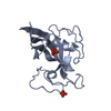 1aqzS S: Starting model for refinement C: citing same article ( |
|---|---|
| Similar structure data |


-Links
 PDBj
PDBj


- Assembly
| Deposited unit | 
| ||||||||
|---|---|---|---|---|---|---|---|---|---|
| 1 | 
| ||||||||
| 2 | 
| ||||||||
| Unit cell |
|
-Components
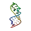
-RNA chain , 3 types, 3 molecules CFD
| #1: RNA chain | Mass: 5809.448 Da / Num. of mol.: 1 / Source method: obtained synthetically |
|---|---|
| #2: RNA chain | Mass: 4234.621 Da / Num. of mol.: 1 / Source method: obtained synthetically |
| #3: RNA chain | Mass: 10027.062 Da / Num. of mol.: 1 / Source method: obtained synthetically Details: The sequence contains 14 highly conserved nucleotides among all living species. |
-Protein , 1 types, 2 molecules AB
| #4: Protein | Mass: 16889.877 Da / Num. of mol.: 2 / Source method: isolated from a natural source / Source: (natural) References: UniProt: P67876, Hydrolases; Acting on ester bonds; Endoribonucleases producing 3'-phosphomonoesters |
|---|
-Non-polymers , 2 types, 173 molecules 

| #5: Chemical | ChemComp-K /  Mass: 39.098 Da / Num. of mol.: 5 / Source method: obtained synthetically / Formula: K Mass: 39.098 Da / Num. of mol.: 5 / Source method: obtained synthetically / Formula: K#6: Water | ChemComp-HOH / | Mass: 18.015 Da / Num. of mol.: 168 / Source method: isolated from a natural source / Formula: H2O |
|---|
-Details
| Has protein modification | Y |
|---|
-Experimental details
-Experiment
| Experiment | Method: X-RAY DIFFRACTION / Number of used crystals: 1 |
|---|
- Sample preparation
| Crystal | Density Matthews: 2.44 Å3/Da / Density % sol: 49.53 % | ||||||||||||||||||||||||||||||||||||||||||
|---|---|---|---|---|---|---|---|---|---|---|---|---|---|---|---|---|---|---|---|---|---|---|---|---|---|---|---|---|---|---|---|---|---|---|---|---|---|---|---|---|---|---|---|
| Crystal grow | Temperature: 292 K / Method: vapor diffusion, hanging drop / pH: 6.5 Details: PEG5000, K-MES, KCl, pH 6.5, VAPOR DIFFUSION, HANGING DROP, temperature 292K | ||||||||||||||||||||||||||||||||||||||||||
| Components of the solutions |
| ||||||||||||||||||||||||||||||||||||||||||
| Crystal grow | *PLUS Temperature: 19 ℃ / pH: 7.2 / Method: vapor diffusion | ||||||||||||||||||||||||||||||||||||||||||
| Components of the solutions | *PLUS
|
-Data collection
| Diffraction | Mean temperature: 100 K |
|---|---|
| Diffraction source | Source: SYNCHROTRON / Site: APS  / Beamline: 14-BM-C / Wavelength: 1 Å / Beamline: 14-BM-C / Wavelength: 1 Å |
| Detector | Type: ADSC QUANTUM 4 / Detector: CCD / Date: Mar 22, 2000 / Details: mirrors |
| Radiation | Monochromator: crystal / Protocol: SINGLE WAVELENGTH / Monochromatic (M) / Laue (L): M / Scattering type: x-ray |
| Radiation wavelength | Wavelength: 1 Å / Relative weight: 1 |
| Reflection | Resolution: 2.15→500 Å / Num. all: 28101 / Num. obs: 24648 / % possible obs: 96.6 % / Observed criterion σ(F): 0 / Observed criterion σ(I): 0 / Redundancy: 2.66 % / Biso Wilson estimate: 51.8 Å2 / Rmerge(I) obs: 0.048 / Net I/σ(I): 24.2 |
| Reflection shell | Resolution: 2.15→2.19 Å / Redundancy: 2.16 % / Rmerge(I) obs: 0.465 / Mean I/σ(I) obs: 2.37 / Num. unique all: 1277 / % possible all: 92.4 |
| Reflection | *PLUS Lowest resolution: 500 Å |
| Reflection shell | *PLUS % possible obs: 92.4 % |
- Processing
| Software |
| ||||||||||||||||||||||||||||||||||||
|---|---|---|---|---|---|---|---|---|---|---|---|---|---|---|---|---|---|---|---|---|---|---|---|---|---|---|---|---|---|---|---|---|---|---|---|---|---|
| Refinement | Method to determine structure: MOLECULAR REPLACEMENT Starting model: PDB entry 1aqz Resolution: 2.15→19.96 Å / Rfactor Rfree error: 0.006 / Isotropic thermal model: isotropic / Cross valid method: THROUGHOUT / σ(F): 0 / σ(I): 0 / Stereochemistry target values: CNS parameter file Details: Electron density around A17 in chain C indicates a mixture of multiple conformations. Only the cleaved conformation has been modeled, and some water molecules around A17 in chain C may ...Details: Electron density around A17 in chain C indicates a mixture of multiple conformations. Only the cleaved conformation has been modeled, and some water molecules around A17 in chain C may represent the alternative conformation(s) of A17.
| ||||||||||||||||||||||||||||||||||||
| Solvent computation | Solvent model: flat model / Bsol: 40.7006 Å2 / ksol: 0.318052 e/Å3 | ||||||||||||||||||||||||||||||||||||
| Displacement parameters | Biso mean: 50.02 Å2
| ||||||||||||||||||||||||||||||||||||
| Refine analyze |
| ||||||||||||||||||||||||||||||||||||
| Refinement step | Cycle: LAST / Resolution: 2.15→19.96 Å
| ||||||||||||||||||||||||||||||||||||
| Refine LS restraints |
| ||||||||||||||||||||||||||||||||||||
| LS refinement shell | Resolution: 2.15→2.28 Å / Rfactor Rfree error: 0.023 / Total num. of bins used: 6
| ||||||||||||||||||||||||||||||||||||
| Xplor file |
| ||||||||||||||||||||||||||||||||||||
| Software | *PLUS Name: CNS / Version: 1 / Classification: refinement | ||||||||||||||||||||||||||||||||||||
| Refinement | *PLUS σ(F): 0 / % reflection Rfree: 9.9 % / Rfactor obs: 0.232 / Rfactor Rfree: 0.29 | ||||||||||||||||||||||||||||||||||||
| Solvent computation | *PLUS | ||||||||||||||||||||||||||||||||||||
| Displacement parameters | *PLUS | ||||||||||||||||||||||||||||||||||||
| Refine LS restraints | *PLUS
| ||||||||||||||||||||||||||||||||||||
| LS refinement shell | *PLUS Rfactor Rfree: 0.426 / % reflection Rfree: 10.5 % / Rfactor Rwork: 0.417 |
