 Movie
Movie Controller


+ Open data

- Basic information
| Entry | Database: PDB / ID: 2o6p | ||||||
|---|---|---|---|---|---|---|---|
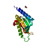
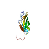
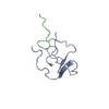
| Title | Crystal Structure of the heme-IsdC complex | ||||||
 Components Components | Iron-regulated surface determinant protein C | ||||||
 Keywords Keywords | TRANSPORT PROTEIN / Beta barrel / protein-heme complex | ||||||
| Function / homology |  Function and homology information Function and homology informationheme transport / hemoglobin binding / peptidoglycan-based cell wall / metal ion binding Similarity search - Function | ||||||
| Biological species |   Staphylococcus aureus subsp. aureus (bacteria) Staphylococcus aureus subsp. aureus (bacteria) | ||||||
| Method |  X-RAY DIFFRACTION / SYNCHROTRON / MAD / Resolution: 1.5 Å X-RAY DIFFRACTION / SYNCHROTRON / MAD / Resolution: 1.5 Å | ||||||
 Authors Authors | Sharp, K.H. / Schneider, S. / Cockayne, A. / Paoli, M. | ||||||
 Citation Citation | Journal: J. Biol. Chem. / Year: 2007 Title: Crystal structure of the heme-IsdC complex, the central conduit of the Isd iron/heme uptake system in Staphylococcus aureus. Authors: Sharp, K.H. / Schneider, S. / Cockayne, A. / Paoli, M. | ||||||
| History |
|
- Structure visualization
| Structure viewer | Molecule: MolmilJmol/JSmol |
|---|
- Downloads & links
-Download
| PDBx/mmCIF format | 2o6p.cif.gz | 125.6 KB | Display | PDBx/mmCIF format |
|---|---|---|---|---|
| PDB format | pdb2o6p.ent.gz | 97.4 KB | Display | PDB format |
| PDBx/mmJSON format | 2o6p.json.gz | Tree view | PDBx/mmJSON format | |
| Others |  Other downloads Other downloads |
-Validation report
| Arichive directory | https://data.pdbj.org/pub/pdb/validation_reports/o6/2o6pftp://data.pdbj.org/pub/pdb/validation_reports/o6/2o6p | HTTPS FTP |
|---|
-Related structure data
| Similar structure data |
|---|


-Links
 PDBj
PDBj- Assembly
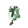
| Deposited unit | 
| ||||||||
|---|---|---|---|---|---|---|---|---|---|
| 1 | 
| ||||||||
| 2 | 
| ||||||||
| 3 |
| ||||||||
| 4 | 
| ||||||||
| 5 | 
| ||||||||
| Unit cell |
|
-Components
| #1: Protein | Mass: 17811.355 Da / Num. of mol.: 2 / Fragment: residues 28-188 Source method: isolated from a genetically manipulated source Source: (gene. exp.) Staphylococcus aureus subsp. aureus (bacteria)Species: Staphylococcus aureus / Strain: N315 / Plasmid: pGAT2 / Species (production host): Escherichia coli / Production host: #2: Chemical | ChemComp-ZN /   Mass: 65.409 Da / Num. of mol.: 6 / Source method: obtained synthetically / Formula: Zn Mass: 65.409 Da / Num. of mol.: 6 / Source method: obtained synthetically / Formula: Zn#3: Chemical |   Mass: 35.453 Da / Num. of mol.: 2 / Source method: obtained synthetically / Formula: Cl Mass: 35.453 Da / Num. of mol.: 2 / Source method: obtained synthetically / Formula: Cl#4: Chemical |   Mass: 616.487 Da / Num. of mol.: 2 / Source method: obtained synthetically / Formula: C34H32FeN4O4 Mass: 616.487 Da / Num. of mol.: 2 / Source method: obtained synthetically / Formula: C34H32FeN4O4#5: Water | ChemComp-HOH / |  Mass: 18.015 Da / Num. of mol.: 189 / Source method: isolated from a natural source / Formula: H2O Mass: 18.015 Da / Num. of mol.: 189 / Source method: isolated from a natural source / Formula: H2O |
|---|
-Experimental details
-Experiment
| Experiment | Method: X-RAY DIFFRACTION / Number of used crystals: 1 |
|---|
- Sample preparation
| Crystal | Density Matthews: 2.08 Å3/Da / Density % sol: 40.91 % |
|---|---|
| Crystal grow | Temperature: 292 K / Method: vapor diffusion, sitting drop / pH: 6 Details: IsdC at 95mg/ml in 50mM Tris-HCl pH 8.0, 500mM NaCl mixed in equal volumes (2 + 2 l) with reservoir solution containing 26% PEG 550, MME, 18mM zinc sulphate, 0.1M MES pH 6.0, VAPOR ...Details: IsdC at 95mg/ml in 50mM Tris-HCl pH 8.0, 500mM NaCl mixed in equal volumes (2 + 2 l) with reservoir solution containing 26% PEG 550, MME, 18mM zinc sulphate, 0.1M MES pH 6.0, VAPOR DIFFUSION, SITTING DROP, temperature 292K |
-Data collection
| Diffraction | Mean temperature: 277 K | |||||||||||||||
|---|---|---|---|---|---|---|---|---|---|---|---|---|---|---|---|---|
| Diffraction source | Source: SYNCHROTRON / Site: ESRF  / Beamline: ID29 / Wavelength: 0.933, 1.739, 1.741, 1.722 / Beamline: ID29 / Wavelength: 0.933, 1.739, 1.741, 1.722 | |||||||||||||||
| Detector | Date: Aug 31, 2006 | |||||||||||||||
| Radiation | Protocol: MAD / Monochromatic (M) / Laue (L): M / Scattering type: x-ray | |||||||||||||||
| Radiation wavelength |
| |||||||||||||||
| Reflection | Resolution: 1.5→47.5 Å / Num. all: 48228 / Num. obs: 45711 / Redundancy: 4.3 % / Rmerge(I) obs: 0.03 / Net I/σ(I): 12.3 | |||||||||||||||
| Reflection shell | Resolution: 1.5→1.6 Å / Redundancy: 4.9 % / Rmerge(I) obs: 0.43 / Mean I/σ(I) obs: 1.8 / Num. unique all: 6918 |
- Processing
| Software |
| |||||||||||||||||||||||||||||||||||||||||||||||||||||||||||||||||||||||||||||||||||||||||||||||||||||||||||||||||||
|---|---|---|---|---|---|---|---|---|---|---|---|---|---|---|---|---|---|---|---|---|---|---|---|---|---|---|---|---|---|---|---|---|---|---|---|---|---|---|---|---|---|---|---|---|---|---|---|---|---|---|---|---|---|---|---|---|---|---|---|---|---|---|---|---|---|---|---|---|---|---|---|---|---|---|---|---|---|---|---|---|---|---|---|---|---|---|---|---|---|---|---|---|---|---|---|---|---|---|---|---|---|---|---|---|---|---|---|---|---|---|---|---|---|---|---|---|
| Refinement | Method to determine structure: MAD / Resolution: 1.5→35.99 Å / Cor.coef. Fo:Fc: 0.962 / Cor.coef. Fo:Fc free: 0.955 / SU B: 3.204 / SU ML: 0.054 / Isotropic thermal model: anisotropic / Cross valid method: THROUGHOUT / ESU R: 0.089 / ESU R Free: 0.076 / Stereochemistry target values: MAXIMUM LIKELIHOOD / Details: HYDROGENS HAVE BEEN ADDED IN THE RIDING POSITIONS
| |||||||||||||||||||||||||||||||||||||||||||||||||||||||||||||||||||||||||||||||||||||||||||||||||||||||||||||||||||
| Solvent computation | Ion probe radii: 0.8 Å / Shrinkage radii: 0.8 Å / VDW probe radii: 1.4 Å / Solvent model: MASK | |||||||||||||||||||||||||||||||||||||||||||||||||||||||||||||||||||||||||||||||||||||||||||||||||||||||||||||||||||
| Displacement parameters | Biso mean: 21.346 Å2
| |||||||||||||||||||||||||||||||||||||||||||||||||||||||||||||||||||||||||||||||||||||||||||||||||||||||||||||||||||
| Refinement step | Cycle: LAST / Resolution: 1.5→35.99 Å
| |||||||||||||||||||||||||||||||||||||||||||||||||||||||||||||||||||||||||||||||||||||||||||||||||||||||||||||||||||
| Refine LS restraints |
| |||||||||||||||||||||||||||||||||||||||||||||||||||||||||||||||||||||||||||||||||||||||||||||||||||||||||||||||||||
| LS refinement shell | Resolution: 1.501→1.54 Å / Total num. of bins used: 20
|
