 Movie
Movie Controller

[English] 日本語
 Yorodumi
- PDB-5f7h: Human T-cell immunoglobulin and mucin domain protein 4 (hTIM-4) c... -
+ Open data

- Basic information
| Entry | Database: PDB / ID: 5f7h | |||||||||
|---|---|---|---|---|---|---|---|---|---|---|
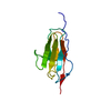
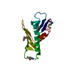
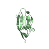
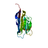
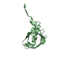
| Title | Human T-cell immunoglobulin and mucin domain protein 4 (hTIM-4) complex with phosphoserine | |||||||||
 Components Components | T-cell immunoglobulin and mucin domain-containing protein 4 | |||||||||
 Keywords Keywords | IMMUNE SYSTEM / complex / hTIM-4 / phosphoserine | |||||||||
| Function / homology |  Function and homology information Function and homology informationcytoskeletal rearrangement involved in phagocytosis, engulfment / Dengue Virus Attachment and Entry / apoptotic cell clearance / phosphatidylserine binding / extracellular region / plasma membrane Similarity search - Function | |||||||||
| Biological species |  Homo sapiens (human) Homo sapiens (human) | |||||||||
| Method |  X-RAY DIFFRACTION / SYNCHROTRON / MOLECULAR REPLACEMENT / Resolution: 2.5 Å X-RAY DIFFRACTION / SYNCHROTRON / MOLECULAR REPLACEMENT / Resolution: 2.5 Å | |||||||||
 Authors Authors | Gao, G.F. / Lu, G. / Wang, H. / Qi, J. | |||||||||
| Funding support |  China, 2items China, 2items
| |||||||||
 Citation Citation | Journal: Chin.Sci.Bull. / Year: 2015 Title: Crystal structures of human TIM members: Ebolavirus entry-enhancing receptors Authors: Wang, H. / Qi, J.X. / Liu, N.N. | |||||||||
| History |
|
- Structure visualization
| Structure viewer | Molecule: MolmilJmol/JSmol |
|---|
- Downloads & links
-Download
| PDBx/mmCIF format | 5f7h.cif.gz | 149.2 KB | Display | PDBx/mmCIF format |
|---|---|---|---|---|
| PDB format | pdb5f7h.ent.gz | 118.1 KB | Display | PDB format |
| PDBx/mmJSON format | 5f7h.json.gz | Tree view | PDBx/mmJSON format | |
| Others |  Other downloads Other downloads |
-Validation report
| Arichive directory | https://data.pdbj.org/pub/pdb/validation_reports/f7/5f7hftp://data.pdbj.org/pub/pdb/validation_reports/f7/5f7h | HTTPS FTP |
|---|
-Related structure data
| Related structure data |  5f70C 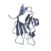 5f71C  5f7fC 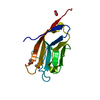 3bi9S C: citing same article ( S: Starting model for refinement |
|---|---|
| Similar structure data |


-Links
 PDBj
PDBj- Assembly
| Deposited unit | 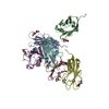
| ||||||||
|---|---|---|---|---|---|---|---|---|---|
| 1 | 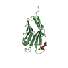
| ||||||||
| 2 | 
| ||||||||
| 3 | 
| ||||||||
| 4 | 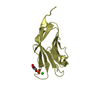
| ||||||||
| 5 | 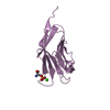
| ||||||||
| 6 | 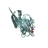
| ||||||||
| Unit cell |
|
-Components
| #1: Protein | Mass: 12667.476 Da / Num. of mol.: 6 / Fragment: UNP residues 24-134 Source method: isolated from a genetically manipulated source Source: (gene. exp.) Homo sapiens (human) / Gene: TIMD4, TIM4 / Production host:  #2: Chemical | ChemComp-CA /   Mass: 40.078 Da / Num. of mol.: 6 / Source method: obtained synthetically / Formula: Ca Mass: 40.078 Da / Num. of mol.: 6 / Source method: obtained synthetically / Formula: Ca#3: Chemical | ChemComp-SEP /   Type: L-peptide linking / Mass: 185.072 Da / Num. of mol.: 6 / Source method: obtained synthetically / Formula: C3H8NO6P Type: L-peptide linking / Mass: 185.072 Da / Num. of mol.: 6 / Source method: obtained synthetically / Formula: C3H8NO6P#4: Water | ChemComp-HOH / |  Mass: 18.015 Da / Num. of mol.: 116 / Source method: isolated from a natural source / Formula: H2O Mass: 18.015 Da / Num. of mol.: 116 / Source method: isolated from a natural source / Formula: H2OHas protein modification | Y | |
|---|
-Experimental details
-Experiment
| Experiment | Method: X-RAY DIFFRACTION |
|---|
- Sample preparation
| Crystal | Density Matthews: 2.97 Å3/Da / Density % sol: 58.54 % |
|---|---|
| Crystal grow | Temperature: 291 K / Method: vapor diffusion, sitting drop Details: hTIM-4 crystals were soaked in reservoir solution with the addition of 20 mM O-Phospho-L-serine (sigma-aldrich) and 5 mM CaCl2 for 20 h. |
-Data collection
| Diffraction | Mean temperature: 100 K |
|---|---|
| Diffraction source | Source: SYNCHROTRON / Site: SSRF / Beamline: BL17U / Wavelength: 0.97915 Å |
| Detector | Type: MARMOSAIC 225 mm CCD / Detector: CCD / Date: May 8, 2015 |
| Radiation | Protocol: SINGLE WAVELENGTH / Monochromatic (M) / Laue (L): M / Scattering type: x-ray |
| Radiation wavelength | Wavelength: 0.97915 Å / Relative weight: 1 |
| Reflection | Resolution: 2.5→50 Å / Num. obs: 30954 / % possible obs: 99.1 % / Redundancy: 4.6 % / Rmerge(I) obs: 0.116 / Rsym value: 0.116 / Net I/σ(I): 12.143 |
| Reflection shell | Resolution: 2.5→2.59 Å / Redundancy: 4.2 % / Rmerge(I) obs: 0.81 / Mean I/σ(I) obs: 1.333 / Rsym value: 0.81 / % possible all: 95.3 |
- Processing
| Software |
| |||||||||||||||||||||||||||||||||||||||||||||||||||||||||||||||||||||||||||||
|---|---|---|---|---|---|---|---|---|---|---|---|---|---|---|---|---|---|---|---|---|---|---|---|---|---|---|---|---|---|---|---|---|---|---|---|---|---|---|---|---|---|---|---|---|---|---|---|---|---|---|---|---|---|---|---|---|---|---|---|---|---|---|---|---|---|---|---|---|---|---|---|---|---|---|---|---|---|---|
| Refinement | Method to determine structure: MOLECULAR REPLACEMENT Starting model: 3BI9 Resolution: 2.5→48.216 Å / SU ML: 0.44 / Cross valid method: FREE R-VALUE / σ(F): 1.35 / Phase error: 32.29 / Stereochemistry target values: ML
| |||||||||||||||||||||||||||||||||||||||||||||||||||||||||||||||||||||||||||||
| Solvent computation | Shrinkage radii: 0.9 Å / VDW probe radii: 1.11 Å / Solvent model: FLAT BULK SOLVENT MODEL | |||||||||||||||||||||||||||||||||||||||||||||||||||||||||||||||||||||||||||||
| Refinement step | Cycle: LAST / Resolution: 2.5→48.216 Å
| |||||||||||||||||||||||||||||||||||||||||||||||||||||||||||||||||||||||||||||
| Refine LS restraints |
| |||||||||||||||||||||||||||||||||||||||||||||||||||||||||||||||||||||||||||||
| LS refinement shell |
|
