 Movie
Movie Controller

[English] 日本語
 Yorodumi
- PDB-1q10: Ensemble of 40 Structures of the Dimeric Mutant of the B1 Domain ... -
+ Open data

- Basic information
| Entry | Database: PDB / ID: 1q10 | ||||||
|---|---|---|---|---|---|---|---|
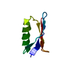
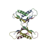
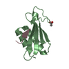
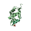
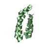
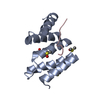
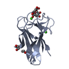
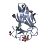
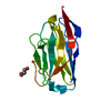
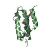
| Title | Ensemble of 40 Structures of the Dimeric Mutant of the B1 Domain of Streptococcal Protein G | ||||||
 Components Components | Immunoglobulin G binding protein G | ||||||
 Keywords Keywords | PROTEIN BINDING / GB1 / core mutants / domain-swapping / oligomerization | ||||||
| Function / homology |  Function and homology information Function and homology information | ||||||
| Biological species |  Streptococcus sp. 'group G' (bacteria) Streptococcus sp. 'group G' (bacteria) | ||||||
| Method | SOLUTION NMR / simulated annealing | ||||||
| Model type details | minimized average | ||||||
 Authors Authors | Byeon, I.J. / Louis, J.M. / Gronenborn, A.M. | ||||||
 Citation Citation | Journal: J.Mol.Biol. / Year: 2003 Title: A Protein Contortionist: Core Mutations of GB1 that Induce Dimerization and Domain Swapping Authors: Byeon, I.J. / Louis, J.M. / Gronenborn, A.M. #1: Journal: Science / Year: 1991Title: A Novel, Highly Stable Fold of the Immunoglobulin Binding Domain of Streptococcal Protein G Authors: Gronenborn, A.M. / Filpula, D.R. / Essig, N.Z. / Achari, A. / Whitlow, M. / Wingfield, P.T. / Clore, G.M. #2: Journal: Nat.Struct.Biol. / Year: 2002Title: Core Mutations Switch Monomeric Protein GB1 Into an Intertwined Tetramer Authors: Frank, M.K. / Dyda, F. / Dobrodumov, A. / Gronenborn, A.M. | ||||||
| History |
|
- Structure visualization
| Structure viewer | Molecule: MolmilJmol/JSmol |
|---|
- Downloads & links
-Download
| PDBx/mmCIF format | 1q10.cif.gz | 1.3 MB | Display | PDBx/mmCIF format |
|---|---|---|---|---|
| PDB format | pdb1q10.ent.gz | 1.1 MB | Display | PDB format |
| PDBx/mmJSON format | 1q10.json.gz | Tree view | PDBx/mmJSON format | |
| Others |  Other downloads Other downloads |
-Validation report
| Arichive directory | https://data.pdbj.org/pub/pdb/validation_reports/q1/1q10ftp://data.pdbj.org/pub/pdb/validation_reports/q1/1q10 | HTTPS FTP |
|---|
-Related structure data
| Related structure data | |
|---|---|
| Similar structure data |


-Links
 PDBj
PDBj

- Assembly
| Deposited unit | 
| |||||||||
|---|---|---|---|---|---|---|---|---|---|---|
| 1 |
| |||||||||
| NMR ensembles |
|
-Components
| #1: Antibody | Mass: 6226.835 Da / Num. of mol.: 2 / Fragment: B1 Domain / Mutation: T228Q,L231V,F256V,Y259F,A260F Source method: isolated from a genetically manipulated source Source: (gene. exp.) Streptococcus sp. 'group G' (bacteria) / Gene: SPG / Plasmid: pET11A / Production host: |
|---|
-Experimental details
-Experiment
| Experiment | Method: SOLUTION NMR | ||||||||||||||||||||||||||||
|---|---|---|---|---|---|---|---|---|---|---|---|---|---|---|---|---|---|---|---|---|---|---|---|---|---|---|---|---|---|
| NMR experiment |
| ||||||||||||||||||||||||||||
| NMR details | Text: The structure was determined using triple-resonance NMR spectroscopy. |
- Sample preparation
| Details |
| ||||||||||||||||||
|---|---|---|---|---|---|---|---|---|---|---|---|---|---|---|---|---|---|---|---|
| Sample conditions | Ionic strength: 50mM sodium phosphate buffer / pH: 5.5 / Pressure: ambient / Temperature: 298 K | ||||||||||||||||||
| Crystal grow | *PLUS Method: other / Details: NMR |
-NMR measurement
| Radiation | Protocol: SINGLE WAVELENGTH / Monochromatic (M) / Laue (L): M | ||||||||||||||||||||||||||||||
|---|---|---|---|---|---|---|---|---|---|---|---|---|---|---|---|---|---|---|---|---|---|---|---|---|---|---|---|---|---|---|---|
| Radiation wavelength | Relative weight: 1 | ||||||||||||||||||||||||||||||
| NMR spectrometer |
|
- Processing
| NMR software |
| ||||||||||||||||||||||||
|---|---|---|---|---|---|---|---|---|---|---|---|---|---|---|---|---|---|---|---|---|---|---|---|---|---|
| Refinement | Method: simulated annealing / Software ordinal: 1 Details: The structures are based on a total of 1311 constraints per monomeric subunit: 1035 NOE-derived distance constraints, 72 hydrogen bond distance constraints, 148 dihedral angle constraints, ...Details: The structures are based on a total of 1311 constraints per monomeric subunit: 1035 NOE-derived distance constraints, 72 hydrogen bond distance constraints, 148 dihedral angle constraints, and 56 residual N-H dipolar coupling constraints. | ||||||||||||||||||||||||
| NMR representative | Selection criteria: minimized average structure | ||||||||||||||||||||||||
| NMR ensemble | Conformer selection criteria: structures with the lowest energy,target function Conformers calculated total number: 100 / Conformers submitted total number: 40 |
 NMRPipe