 Movie
Movie Controller


+ Open data

- Basic information
| Entry | Database: PDB / ID: 3jzq | ||||||
|---|---|---|---|---|---|---|---|
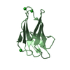
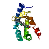
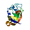
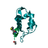
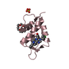
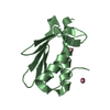
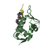
| Title | Human MDMX liganded with a 12mer peptide inhibitor (pDIQ) | ||||||
 Components Components |
| ||||||
 Keywords Keywords | CELL CYCLE / P53-BINDING PROTEIN MDM4 / DOUBLE MINUTE 4 PROTEIN / Alternative splicing / Metal-binding / Nucleus / Polymorphism / Zinc / Zinc-finger | ||||||
| Function / homology |  Function and homology information Function and homology informationatrial septum development / heart valve development / atrioventricular valve morphogenesis / ventricular septum development / endocardial cushion morphogenesis / negative regulation of signal transduction by p53 class mediator / transcription repressor complex / DNA damage response, signal transduction by p53 class mediator / Stabilization of p53 / negative regulation of protein catabolic process ...atrial septum development / heart valve development / atrioventricular valve morphogenesis / ventricular septum development / endocardial cushion morphogenesis / negative regulation of signal transduction by p53 class mediator / transcription repressor complex / DNA damage response, signal transduction by p53 class mediator / Stabilization of p53 / negative regulation of protein catabolic process / Oncogene Induced Senescence / Regulation of TP53 Activity through Methylation / enzyme activator activity / ubiquitin-protein transferase activity / Regulation of TP53 Degradation / protein-containing complex assembly / Oxidative Stress Induced Senescence / cellular response to hypoxia / Regulation of TP53 Activity through Phosphorylation / regulation of cell cycle / protein stabilization / Ub-specific processing proteases / protein ubiquitination / negative regulation of cell population proliferation / negative regulation of DNA-templated transcription / negative regulation of apoptotic process / enzyme binding / negative regulation of transcription by RNA polymerase II / zinc ion binding / nucleoplasm / nucleus Similarity search - Function | ||||||
| Biological species |  Homo sapiens (human) Homo sapiens (human) | ||||||
| Method |  X-RAY DIFFRACTION / MOLECULAR REPLACEMENT / Resolution: 1.8 Å X-RAY DIFFRACTION / MOLECULAR REPLACEMENT / Resolution: 1.8 Å | ||||||
 Authors Authors | Schonbrunn, E. / Phan, J. | ||||||
 Citation Citation | Journal: J.Biol.Chem. / Year: 2010 Title: Structure-based design of high affinity peptides inhibiting the interaction of p53 with MDM2 and MDMX. Authors: Phan, J. / Li, Z. / Kasprzak, A. / Li, B. / Sebti, S. / Guida, W. / Schonbrunn, E. / Chen, J. | ||||||
| History |
|
- Structure visualization
| Structure viewer | Molecule: MolmilJmol/JSmol |
|---|
- Downloads & links
-Download
| PDBx/mmCIF format | 3jzq.cif.gz | 54.6 KB | Display | PDBx/mmCIF format |
|---|---|---|---|---|
| PDB format | pdb3jzq.ent.gz | 39.4 KB | Display | PDB format |
| PDBx/mmJSON format | 3jzq.json.gz | Tree view | PDBx/mmJSON format | |
| Others |  Other downloads Other downloads |
-Validation report
| Arichive directory | https://data.pdbj.org/pub/pdb/validation_reports/jz/3jzqftp://data.pdbj.org/pub/pdb/validation_reports/jz/3jzq | HTTPS FTP |
|---|
-Related structure data
| Related structure data |  3jzoC  3jzpC  3jzrC  3jzsC 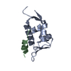 3dabS C: citing same article ( S: Starting model for refinement |
|---|---|
| Similar structure data |


-Links
 PDBj
PDBj

- Assembly
| Deposited unit | 
| ||||||||
|---|---|---|---|---|---|---|---|---|---|
| 1 | 
| ||||||||
| 2 | 
| ||||||||
| Unit cell |
|
-Components
| #1: Protein | Mass: 10160.899 Da / Num. of mol.: 2 Source method: isolated from a genetically manipulated source Source: (gene. exp.) Homo sapiens (human) / Gene: MDM4, MDMX / Plasmid: pDEST-His-MBP / Production host:  #2: Protein/peptide | Mass: 1563.687 Da / Num. of mol.: 2 / Source method: obtained synthetically #3: Chemical | ChemComp-SO4 / |   Mass: 96.063 Da / Num. of mol.: 1 / Source method: obtained synthetically / Formula: SO4 Mass: 96.063 Da / Num. of mol.: 1 / Source method: obtained synthetically / Formula: SO4#4: Water | ChemComp-HOH / |  Mass: 18.015 Da / Num. of mol.: 139 / Source method: isolated from a natural source / Formula: H2O Mass: 18.015 Da / Num. of mol.: 139 / Source method: isolated from a natural source / Formula: H2O |
|---|
-Experimental details
-Experiment
| Experiment | Method: X-RAY DIFFRACTION / Number of used crystals: 1 |
|---|
- Sample preparation
| Crystal | Density Matthews: 2.25 Å3/Da / Density % sol: 45.25 % |
|---|---|
| Crystal grow | Temperature: 292 K / Method: vapor diffusion, hanging drop / pH: 7.4 Details: 2.1 M ammonium sulfate, 10 mM Tris HCl, pH 7.4, VAPOR DIFFUSION, HANGING DROP, temperature 292K |
-Data collection
| Diffraction | Mean temperature: 93 K |
|---|---|
| Diffraction source | Source: ROTATING ANODE / Type: RIGAKU MICROMAX-007 HF / Wavelength: 1.54 Å |
| Detector | Type: RIGAKU RAXIS HTC / Detector: IMAGE PLATE / Details: mirrors |
| Radiation | Monochromator: mirrors / Protocol: SINGLE WAVELENGTH / Monochromatic (M) / Laue (L): M / Scattering type: x-ray |
| Radiation wavelength | Wavelength: 1.54 Å / Relative weight: 1 |
| Reflection | Resolution: 1.8→50 Å / Num. all: 20193 / Num. obs: 20193 / % possible obs: 99.7 % / Observed criterion σ(F): 0 / Observed criterion σ(I): 0 / Redundancy: 6.9 % / Biso Wilson estimate: 25.7 Å2 / Rmerge(I) obs: 0.062 / Net I/σ(I): 27.5 |
| Reflection shell | Resolution: 1.8→1.86 Å / Redundancy: 6.7 % / Rmerge(I) obs: 0.47 / Mean I/σ(I) obs: 3.7 / Num. unique all: 1953 / % possible all: 99 |
- Processing
| Software |
| |||||||||||||||||||||||||
|---|---|---|---|---|---|---|---|---|---|---|---|---|---|---|---|---|---|---|---|---|---|---|---|---|---|---|
| Refinement | Method to determine structure: MOLECULAR REPLACEMENT Starting model: pdb entry 3DAB Resolution: 1.8→34.5 Å / Cross valid method: THROUGHOUT / σ(F): 0 / σ(I): 0 / Stereochemistry target values: Engh & Huber
| |||||||||||||||||||||||||
| Refine analyze |
| |||||||||||||||||||||||||
| Refinement step | Cycle: LAST / Resolution: 1.8→34.5 Å
| |||||||||||||||||||||||||
| Refine LS restraints |
| |||||||||||||||||||||||||
| LS refinement shell | Resolution: 1.8→1.91 Å / Rfactor Rfree error: 0.03
|
