 Movie
Movie Controller


+ Open data

- Basic information
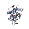
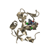
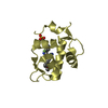
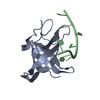
| Entry | Database: PDB / ID: 3jzr | ||||||
|---|---|---|---|---|---|---|---|
| Title | Human MDM2 liganded with a 12mer peptide inhibitor (pDI6W) | ||||||
 Components Components |
| ||||||
 Keywords Keywords | LIGASE / P53-BINDING PROTEIN MDM2 / ONCOPROTEIN MDM2 / DOUBLE MINUTE 2 PROTEIN / HDM2 / Alternative splicing / Cytoplasm / Host-virus interaction / Metal-binding / Nucleus / Phosphoprotein / Proto-oncogene / Ubl conjugation / Ubl conjugation pathway / Zinc / Zinc-finger | ||||||
| Function / homology |  Function and homology information Function and homology informationcellular response to vitamin B1 / response to formaldehyde / response to water-immersion restraint stress / response to ether / traversing start control point of mitotic cell cycle / atrial septum development / fibroblast activation / regulation of protein catabolic process at postsynapse, modulating synaptic transmission / Trafficking of AMPA receptors / receptor serine/threonine kinase binding ...cellular response to vitamin B1 / response to formaldehyde / response to water-immersion restraint stress / response to ether / traversing start control point of mitotic cell cycle / atrial septum development / fibroblast activation / regulation of protein catabolic process at postsynapse, modulating synaptic transmission / Trafficking of AMPA receptors / receptor serine/threonine kinase binding / negative regulation of intrinsic apoptotic signaling pathway by p53 class mediator / negative regulation of protein processing / response to steroid hormone / SUMO transferase activity / peroxisome proliferator activated receptor binding / positive regulation of vascular associated smooth muscle cell migration / atrioventricular valve morphogenesis / AKT phosphorylates targets in the cytosol / response to iron ion / NEDD8 ligase activity / endocardial cushion morphogenesis / cellular response to peptide hormone stimulus / ventricular septum development / positive regulation of muscle cell differentiation / cardiac septum morphogenesis / regulation of postsynaptic neurotransmitter receptor internalization / SUMOylation of ubiquitinylation proteins / cellular response to alkaloid / blood vessel development / ligase activity / Constitutive Signaling by AKT1 E17K in Cancer / negative regulation of DNA damage response, signal transduction by p53 class mediator / cellular response to antibiotic / SUMOylation of transcription factors / negative regulation of signal transduction by p53 class mediator / regulation of protein catabolic process / cellular response to UV-C / cellular response to estrogen stimulus / protein sumoylation / response to magnesium ion / blood vessel remodeling / ribonucleoprotein complex binding / protein localization to nucleus / protein autoubiquitination / positive regulation of vascular associated smooth muscle cell proliferation / NPAS4 regulates expression of target genes / transcription repressor complex / positive regulation of mitotic cell cycle / regulation of heart rate / : / positive regulation of protein export from nucleus / response to cocaine / ubiquitin binding / DNA damage response, signal transduction by p53 class mediator / establishment of protein localization / sperm end piece / Stabilization of p53 / cellular response to gamma radiation / Regulation of RUNX3 expression and activity / RING-type E3 ubiquitin transferase / Oncogene Induced Senescence / Regulation of TP53 Activity through Methylation / Degradation of CDH1 / protein destabilization / cellular response to growth factor stimulus / response to toxic substance / cellular response to hydrogen peroxide / centriolar satellite / disordered domain specific binding / protein polyubiquitination / p53 binding / ubiquitin-protein transferase activity / endocytic vesicle membrane / Signaling by ALK fusions and activated point mutants / Regulation of TP53 Degradation / ubiquitin protein ligase activity / negative regulation of neuron projection development / positive regulation of proteasomal ubiquitin-dependent protein catabolic process / sperm principal piece / 5S rRNA binding / protein-containing complex assembly / sperm midpiece / Oxidative Stress Induced Senescence / cellular response to hypoxia / Regulation of TP53 Activity through Phosphorylation / amyloid fibril formation / ubiquitin-dependent protein catabolic process / proteasome-mediated ubiquitin-dependent protein catabolic process / regulation of cell cycle / postsynaptic density / Ub-specific processing proteases / protein ubiquitination / response to xenobiotic stimulus / protein domain specific binding / response to antibiotic / negative regulation of DNA-templated transcription / apoptotic process / positive regulation of cell population proliferation / ubiquitin protein ligase binding / positive regulation of gene expression Similarity search - Function | ||||||
| Biological species |  Homo sapiens (human) Homo sapiens (human) | ||||||
| Method |  X-RAY DIFFRACTION / MOLECULAR REPLACEMENT / Resolution: 2.1 Å X-RAY DIFFRACTION / MOLECULAR REPLACEMENT / Resolution: 2.1 Å | ||||||
 Authors Authors | Schonbrunn, E. / Phan, J. | ||||||
 Citation Citation | Journal: J.Biol.Chem. / Year: 2010 Title: Structure-based design of high affinity peptides inhibiting the interaction of p53 with MDM2 and MDMX. Authors: Phan, J. / Li, Z. / Kasprzak, A. / Li, B. / Sebti, S. / Guida, W. / Schonbrunn, E. / Chen, J. | ||||||
| History |
|
- Structure visualization
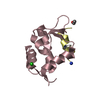
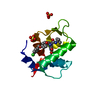
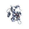
| Structure viewer | Molecule: MolmilJmol/JSmol |
|---|
- Downloads & links
-Download
| PDBx/mmCIF format | 3jzr.cif.gz | 37.8 KB | Display | PDBx/mmCIF format |
|---|---|---|---|---|
| PDB format | pdb3jzr.ent.gz | 25 KB | Display | PDB format |
| PDBx/mmJSON format | 3jzr.json.gz | Tree view | PDBx/mmJSON format | |
| Others |  Other downloads Other downloads |
-Validation report
| Arichive directory | https://data.pdbj.org/pub/pdb/validation_reports/jz/3jzrftp://data.pdbj.org/pub/pdb/validation_reports/jz/3jzr | HTTPS FTP |
|---|
-Related structure data
| Related structure data |  3jzoC  3jzpC  3jzqC  3jzsC  1t4fS C: citing same article ( S: Starting model for refinement |
|---|---|
| Similar structure data |


-Links
 PDBj
PDBj


- Assembly
| Deposited unit | 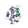
| ||||||||
|---|---|---|---|---|---|---|---|---|---|
| 1 |
| ||||||||
| Unit cell |
|
-Components
| #1: Protein | Mass: 12592.401 Da / Num. of mol.: 1 Source method: isolated from a genetically manipulated source Source: (gene. exp.) Homo sapiens (human) / Gene: MDM2 / Plasmid: pGEX2T / Production host:  References: UniProt: Q00987, Ligases; Forming carbon-nitrogen bonds; Acid-amino-acid ligases (peptide synthases) |
|---|---|
| #2: Protein/peptide | Mass: 1519.677 Da / Num. of mol.: 1 / Source method: obtained synthetically |
| #3: Water | ChemComp-HOH /  Mass: 18.015 Da / Num. of mol.: 116 / Source method: isolated from a natural source / Formula: H2O Mass: 18.015 Da / Num. of mol.: 116 / Source method: isolated from a natural source / Formula: H2O |
-Experimental details
-Experiment
| Experiment | Method: X-RAY DIFFRACTION / Number of used crystals: 1 |
|---|
- Sample preparation
| Crystal | Density Matthews: 2.1 Å3/Da / Density % sol: 41.34 % |
|---|---|
| Crystal grow | Temperature: 292 K / Method: vapor diffusion, hanging drop / pH: 8 Details: 2.2 M DL-Malic acid, 100 mM Tris HCl, pH 8.0, VAPOR DIFFUSION, HANGING DROP, temperature 292K |
-Data collection
| Diffraction | Mean temperature: 93 K |
|---|---|
| Diffraction source | Source: ROTATING ANODE / Type: RIGAKU MICROMAX-007 HF / Wavelength: 1.54 Å |
| Detector | Type: RIGAKU RAXIS HTC / Detector: IMAGE PLATE / Details: mirrors |
| Radiation | Monochromator: mirrors / Protocol: SINGLE WAVELENGTH / Monochromatic (M) / Laue (L): M / Scattering type: x-ray |
| Radiation wavelength | Wavelength: 1.54 Å / Relative weight: 1 |
| Reflection | Resolution: 2.1→50 Å / Num. all: 7261 / Num. obs: 7261 / % possible obs: 99.8 % / Observed criterion σ(F): 0 / Observed criterion σ(I): 0 / Redundancy: 4.4 % / Biso Wilson estimate: 26.6 Å2 / Rmerge(I) obs: 0.12 / Net I/σ(I): 30.1 |
| Reflection shell | Resolution: 2.1→2.18 Å / Redundancy: 4.4 % / Rmerge(I) obs: 0.32 / Mean I/σ(I) obs: 8.4 / Num. unique all: 714 / % possible all: 100 |
- Processing
| Software |
| |||||||||||||||||||||||||
|---|---|---|---|---|---|---|---|---|---|---|---|---|---|---|---|---|---|---|---|---|---|---|---|---|---|---|
| Refinement | Method to determine structure: MOLECULAR REPLACEMENT Starting model: pdb entry 1T4F Resolution: 2.1→46 Å / Cross valid method: THROUGHOUT / σ(F): 0 / σ(I): 0 / Stereochemistry target values: Engh & Huber
| |||||||||||||||||||||||||
| Refine analyze |
| |||||||||||||||||||||||||
| Refinement step | Cycle: LAST / Resolution: 2.1→46 Å
| |||||||||||||||||||||||||
| Refine LS restraints |
| |||||||||||||||||||||||||
| LS refinement shell | Resolution: 2.1→2.23 Å / Rfactor Rfree error: 0.032
|
