 Movie
Movie Controller

[English] 日本語
 Yorodumi
- PDB-1mpe: Ensemble of 20 structures of the tetrameric mutant of the B1 doma... -
+ Open data

- Basic information
| Entry | Database: PDB / ID: 1mpe | ||||||
|---|---|---|---|---|---|---|---|
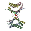
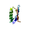
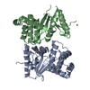
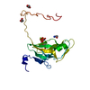
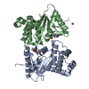
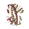
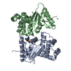
| Title | Ensemble of 20 structures of the tetrameric mutant of the B1 domain of streptococcal protein G | ||||||
 Components Components | Immunoglobulin G binding protein G | ||||||
 Keywords Keywords | PROTEIN BINDING / strand-exchanged tetramer / channel | ||||||
| Function / homology |  Function and homology information Function and homology information | ||||||
| Biological species |  Streptococcus sp. 'group G' (bacteria) Streptococcus sp. 'group G' (bacteria) | ||||||
| Method | SOLUTION NMR / simulated annealing | ||||||
| Model type details | minimized average | ||||||
 Authors Authors | Frank, M.K. / Dyda, F. / Dobrodumov, A. / Gronenborn, A.M. | ||||||
 Citation Citation | Journal: Nat.Struct.Biol. / Year: 2002 Title: Core mutations switch monomeric protein GB1 into an intertwined tetramer. Authors: Kirsten Frank, M. / Dyda, F. / Dobrodumov, A. / Gronenborn, A.M. #1: Journal: Science / Year: 1991Title: A Novel, Highly Stable Fold of the Immunoglobulin Binding Domain of Streptococcal Protein G. Authors: Gronenborn, A.M. / Filpula, D.R. / Essig, N.Z. / Achari, A. / Whitlow, M. / Wingfield, P.T. / Clore, G.M. #2: Journal: FEBS Lett. / Year: 1996Title: Core Mutants of the Immunoglobulin Binding Domain of Streptococcal Protein G: Stability and Structural Integrity Authors: Gronenborn, A.M. / Frank, M.K. / Clore, G.M. | ||||||
| History |
|
- Structure visualization
| Structure viewer | Molecule: MolmilJmol/JSmol |
|---|
- Downloads & links
-Download
| PDBx/mmCIF format | 1mpe.cif.gz | 1.4 MB | Display | PDBx/mmCIF format |
|---|---|---|---|---|
| PDB format | pdb1mpe.ent.gz | 1.2 MB | Display | PDB format |
| PDBx/mmJSON format | 1mpe.json.gz | Tree view | PDBx/mmJSON format | |
| Others |  Other downloads Other downloads |
-Validation report
| Arichive directory | https://data.pdbj.org/pub/pdb/validation_reports/mp/1mpeftp://data.pdbj.org/pub/pdb/validation_reports/mp/1mpe | HTTPS FTP |
|---|
-Related structure data


-Links
 PDBj
PDBj

- Assembly
| Deposited unit | 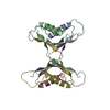
| |||||||||
|---|---|---|---|---|---|---|---|---|---|---|
| 1 |
| |||||||||
| NMR ensembles |
|
-Components
| #1: Antibody | Mass: 6302.931 Da / Num. of mol.: 4 / Fragment: B1 domain, sequence database residues 228-282 / Mutation: T2Q, L5V, A26F, F30V, Y33F, A34F Source method: isolated from a genetically manipulated source Source: (gene. exp.) Streptococcus sp. 'group G' (bacteria) / Gene: SPG / Plasmid: pet11a / Production host: |
|---|
-Experimental details
-Experiment
| Experiment | Method: SOLUTION NMR | ||||||||||||||||||||||||||||
|---|---|---|---|---|---|---|---|---|---|---|---|---|---|---|---|---|---|---|---|---|---|---|---|---|---|---|---|---|---|
| NMR experiment |
| ||||||||||||||||||||||||||||
| NMR details | Text: T1,T2, and heteronuclear NOEs collected. Dipolar couplings (N-H) were collected as well. Dipolar coupling and chemical shifts from residues with heteronuclear NOE below 0.6 were not used. |
- Sample preparation
| Details |
| |||||||||||||||
|---|---|---|---|---|---|---|---|---|---|---|---|---|---|---|---|---|
| Sample conditions | Ionic strength: 50 mM sodium phosphate, 0.02% sodium azide / pH: 5.45 / Pressure: 1 atm / Temperature: 313 K | |||||||||||||||
| Crystal grow | *PLUS Method: other / Details: NMR |
-NMR measurement
| Radiation | Protocol: SINGLE WAVELENGTH / Monochromatic (M) / Laue (L): M | |||||||||||||||||||||||||
|---|---|---|---|---|---|---|---|---|---|---|---|---|---|---|---|---|---|---|---|---|---|---|---|---|---|---|
| Radiation wavelength | Relative weight: 1 | |||||||||||||||||||||||||
| NMR spectrometer |
|
- Processing
| NMR software |
| ||||||||||||||||||||||||
|---|---|---|---|---|---|---|---|---|---|---|---|---|---|---|---|---|---|---|---|---|---|---|---|---|---|
| Refinement | Method: simulated annealing / Software ordinal: 1 Details: NMR constraints (per monomer) 822 NOE-derived distance restraints (534 intramonomer, 288 intermonomer) 42 distance restraints from hydrogen bonds (20 intramonomer, 22 intermonomer) 30 J- ...Details: NMR constraints (per monomer) 822 NOE-derived distance restraints (534 intramonomer, 288 intermonomer) 42 distance restraints from hydrogen bonds (20 intramonomer, 22 intermonomer) 30 J-coupling restraints, 33 Residual dipolar couplings (HN) 66 backbone dihedral angle restraints 31 sidechain dihedral angle restraints | ||||||||||||||||||||||||
| NMR representative | Selection criteria: minimized average structure | ||||||||||||||||||||||||
| NMR ensemble | Conformer selection criteria: structures with least restraint violations and phi, psi angles of residue 46 in allowed region of Ramachrandran plot Conformers calculated total number: 100 / Conformers submitted total number: 21 |
 NMRPipe