 Movie
Movie Controller

[English] 日本語
 Yorodumi
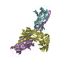
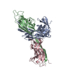
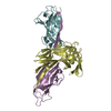
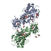
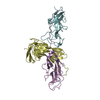
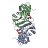
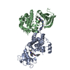
- PDB-1p5u: X-ray structure of the ternary Caf1M:Caf1:Caf1 chaperone:subunit:... -
+ Open data

- Basic information
| Entry | Database: PDB / ID: 1p5u | ||||||
|---|---|---|---|---|---|---|---|
| Title | X-ray structure of the ternary Caf1M:Caf1:Caf1 chaperone:subunit:subunit complex | ||||||
 Components Components |
| ||||||
 Keywords Keywords | CHAPERONE / STRUCTURAL PROTEIN / chaperone-target complex / chaperone-subunit complex / protein fiber / donor strand complementation / donor strand exchange | ||||||
| Function / homology |  Function and homology information Function and homology informationcapsule / pilus / protein folding chaperone / cell wall organization / outer membrane-bounded periplasmic space / cell adhesion Similarity search - Function | ||||||
| Biological species |   Yersinia pestis (bacteria) Yersinia pestis (bacteria) | ||||||
| Method |  X-RAY DIFFRACTION / SYNCHROTRON / MOLECULAR REPLACEMENT / Resolution: 1.99 Å X-RAY DIFFRACTION / SYNCHROTRON / MOLECULAR REPLACEMENT / Resolution: 1.99 Å | ||||||
 Authors Authors | Zavialov, A.V. / Berglund, J. / Pudney, A.F. / Fooks, L.J. / Ibrahim, T.M. / MacIntyre, S. / Knight, S.D. | ||||||
 Citation Citation | Journal: Cell(Cambridge,Mass.) / Year: 2003 Title: Structure and Biogenesis of the Capsular F1 Antigen from Yersinia pestis. Preserved Folding Energy Drives Fiber Formation Authors: Zavialov, A.V. / Berglund, J. / Pudney, A.F. / Fooks, L.J. / Ibrahim, T.M. / MacIntyre, S. / Knight, S.D. #1: Journal: Acta Crystallogr.,Sect.D / Year: 2003Title: Overexpression, purification, crystallization and preliminary X-ray diffraction analysis of the F1 antigen Caf1MCaf1 chaperonesubunit pre-assembly complex from Yersinia pestis Authors: Zavialov, A. / Berglund, J. / Knight, S.D. #2: Journal: Mol.Microbiol. / Year: 2002Title: Donor strand complementation mechanism in the biogenesis of non-pilus systems Authors: Zavialov, A.V. / Kersley, J. / Korpela, T. / Zav'yalov, V.P. / MacIntyre, S. / Knight, S.D. | ||||||
| History |
|
- Structure visualization
| Structure viewer | Molecule: MolmilJmol/JSmol |
|---|
- Downloads & links
-Download
| PDBx/mmCIF format | 1p5u.cif.gz | 113 KB | Display | PDBx/mmCIF format |
|---|---|---|---|---|
| PDB format | pdb1p5u.ent.gz | 86.3 KB | Display | PDB format |
| PDBx/mmJSON format | 1p5u.json.gz | Tree view | PDBx/mmJSON format | |
| Others |  Other downloads Other downloads |
-Validation report
| Arichive directory | https://data.pdbj.org/pub/pdb/validation_reports/p5/1p5uftp://data.pdbj.org/pub/pdb/validation_reports/p5/1p5u | HTTPS FTP |
|---|
-Related structure data


-Links
 PDBj
PDBj
- Assembly
| Deposited unit | 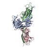
| ||||||||||
|---|---|---|---|---|---|---|---|---|---|---|---|
| 1 |
| ||||||||||
| Unit cell |
|
-Components
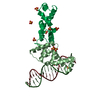
| #1: Protein | Mass: 26329.012 Da / Num. of mol.: 1 / Fragment: residues 24-258 of SWS P26926 Source method: isolated from a genetically manipulated source Source: (gene. exp.) Yersinia pestis (bacteria) / Strain: KIM5 / Gene: CAF1M / Plasmid: pFM-1-6H / Production host: |
|---|---|
| #2: Protein | Mass: 15661.271 Da / Num. of mol.: 1 / Fragment: residues 22-170 of SWS P26948 / Mutation: A9R Source method: isolated from a genetically manipulated source Details: larger fragment of F1 capsule with A9R mutation / Source: (gene. exp.) Yersinia pestis (bacteria) / Strain: KIM5 / Gene: CAF1 / Plasmid: pFM-1-6H / Production host: |
| #3: Protein | Mass: 15673.230 Da / Num. of mol.: 1 / Fragment: residues 35-170 of SWS P26948 Source method: isolated from a genetically manipulated source Details: smaller HIS-tagged fragment of F1 capsule / Source: (gene. exp.) Yersinia pestis (bacteria) / Strain: KIM5 / Gene: CAF1 / Plasmid: pFM-1-6H / Production host: |
| #4: Water | ChemComp-HOH /  Mass: 18.015 Da / Num. of mol.: 290 / Source method: isolated from a natural source / Formula: H2O Mass: 18.015 Da / Num. of mol.: 290 / Source method: isolated from a natural source / Formula: H2O |
| Has protein modification | Y |
-Experimental details
-Experiment
| Experiment | Method: X-RAY DIFFRACTION / Number of used crystals: 1 |
|---|
- Sample preparation
| Crystal | Density Matthews: 2.44 Å3/Da / Density % sol: 49.64 % | ||||||||||||||||||||||||||||||
|---|---|---|---|---|---|---|---|---|---|---|---|---|---|---|---|---|---|---|---|---|---|---|---|---|---|---|---|---|---|---|---|
| Crystal grow | Temperature: 293 K / Method: vapor diffusion, hanging drop / pH: 5.9 Details: PEG 8000, calcium acetate, cacodylate, pH 5.9, VAPOR DIFFUSION, HANGING DROP, temperature 293K | ||||||||||||||||||||||||||||||
| Crystal grow | *PLUS Temperature: 293 K / Method: vapor diffusion, hanging drop | ||||||||||||||||||||||||||||||
| Components of the solutions | *PLUS
|
-Data collection
| Diffraction | Mean temperature: 100 K |
|---|---|
| Diffraction source | Source: SYNCHROTRON / Site: ESRF  / Beamline: ID14-2 / Wavelength: 0.933 Å / Beamline: ID14-2 / Wavelength: 0.933 Å |
| Detector | Type: ADSC QUANTUM 4 / Detector: CCD / Date: Sep 29, 2002 |
| Radiation | Monochromator: Diamond (111), Ge(220) / Protocol: SINGLE WAVELENGTH / Monochromatic (M) / Laue (L): M / Scattering type: x-ray |
| Radiation wavelength | Wavelength: 0.933 Å / Relative weight: 1 |
| Reflection | Resolution: 1.99→87.71 Å / Num. all: 34536 / Num. obs: 34536 / Observed criterion σ(F): 0 / Observed criterion σ(I): 0 / Redundancy: 5.4 % / Biso Wilson estimate: 23.9 Å2 / Rmerge(I) obs: 0.086 / Net I/σ(I): 5.4 |
| Reflection shell | Resolution: 1.99→2.15 Å / Redundancy: 3.4 % / Rmerge(I) obs: 0.24 / Mean I/σ(I) obs: 3 / % possible all: 82 |
| Reflection | *PLUS % possible obs: 88.6 % |
| Reflection shell | *PLUS % possible obs: 82 % |
- Processing
| Software |
| ||||||||||||||||||||||||||||||||||||||||||||||||||||||||||||||||||||||||||||||||||||||||||||||||||||||||||||||||||||||||||||||||||
|---|---|---|---|---|---|---|---|---|---|---|---|---|---|---|---|---|---|---|---|---|---|---|---|---|---|---|---|---|---|---|---|---|---|---|---|---|---|---|---|---|---|---|---|---|---|---|---|---|---|---|---|---|---|---|---|---|---|---|---|---|---|---|---|---|---|---|---|---|---|---|---|---|---|---|---|---|---|---|---|---|---|---|---|---|---|---|---|---|---|---|---|---|---|---|---|---|---|---|---|---|---|---|---|---|---|---|---|---|---|---|---|---|---|---|---|---|---|---|---|---|---|---|---|---|---|---|---|---|---|---|---|
| Refinement | Method to determine structure: MOLECULAR REPLACEMENT Starting model: Refined model of binary Caf1M:Caf1 pre-assembly complex Resolution: 1.99→87.71 Å / Cor.coef. Fo:Fc: 0.958 / Cor.coef. Fo:Fc free: 0.942 / SU B: 3.625 / SU ML: 0.101 / Cross valid method: THROUGHOUT / ESU R: 0.171 / ESU R Free: 0.16 / Stereochemistry target values: MAXIMUM LIKELIHOOD / Details: HYDROGENS HAVE BEEN ADDED IN THE
| ||||||||||||||||||||||||||||||||||||||||||||||||||||||||||||||||||||||||||||||||||||||||||||||||||||||||||||||||||||||||||||||||||
| Solvent computation | Ion probe radii: 0.8 Å / Shrinkage radii: 0.8 Å / VDW probe radii: 1.4 Å / Solvent model: BABINET MODEL WITH MASK | ||||||||||||||||||||||||||||||||||||||||||||||||||||||||||||||||||||||||||||||||||||||||||||||||||||||||||||||||||||||||||||||||||
| Displacement parameters | Biso mean: 17.994 Å2
| ||||||||||||||||||||||||||||||||||||||||||||||||||||||||||||||||||||||||||||||||||||||||||||||||||||||||||||||||||||||||||||||||||
| Refine analyze |
| ||||||||||||||||||||||||||||||||||||||||||||||||||||||||||||||||||||||||||||||||||||||||||||||||||||||||||||||||||||||||||||||||||
| Refinement step | Cycle: LAST / Resolution: 1.99→87.71 Å
| ||||||||||||||||||||||||||||||||||||||||||||||||||||||||||||||||||||||||||||||||||||||||||||||||||||||||||||||||||||||||||||||||||
| Refine LS restraints |
| ||||||||||||||||||||||||||||||||||||||||||||||||||||||||||||||||||||||||||||||||||||||||||||||||||||||||||||||||||||||||||||||||||
| LS refinement shell | Resolution: 1.995→2.046 Å / Total num. of bins used: 20 /
| ||||||||||||||||||||||||||||||||||||||||||||||||||||||||||||||||||||||||||||||||||||||||||||||||||||||||||||||||||||||||||||||||||
| Refinement TLS params. | Method: refined / Refine-ID: X-RAY DIFFRACTION
| ||||||||||||||||||||||||||||||||||||||||||||||||||||||||||||||||||||||||||||||||||||||||||||||||||||||||||||||||||||||||||||||||||
| Refinement TLS group |
| ||||||||||||||||||||||||||||||||||||||||||||||||||||||||||||||||||||||||||||||||||||||||||||||||||||||||||||||||||||||||||||||||||
| Refinement | *PLUS Highest resolution: 2 Å / Lowest resolution: 20 Å / Num. reflection Rfree: 1753 / Rfactor Rfree: 0.225 / Rfactor Rwork: 0.169 | ||||||||||||||||||||||||||||||||||||||||||||||||||||||||||||||||||||||||||||||||||||||||||||||||||||||||||||||||||||||||||||||||||
| Solvent computation | *PLUS | ||||||||||||||||||||||||||||||||||||||||||||||||||||||||||||||||||||||||||||||||||||||||||||||||||||||||||||||||||||||||||||||||||
| Displacement parameters | *PLUS | ||||||||||||||||||||||||||||||||||||||||||||||||||||||||||||||||||||||||||||||||||||||||||||||||||||||||||||||||||||||||||||||||||
| Refine LS restraints | *PLUS
|
