 Movie
Movie Controller

[English] 日本語
 Yorodumi
- PDB-3dsn: Crystal structure of the complex of the Caf1M chaperone with the ... -
+ Open data

- Basic information
| Entry | Database: PDB / ID: 3dsn | ||||||
|---|---|---|---|---|---|---|---|
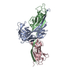
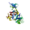
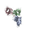
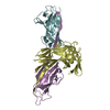
| Title | Crystal structure of the complex of the Caf1M chaperone with the mini-fiber of two Caf1 subunits (Caf1:Caf1), carrying the Thr7Phe mutation in the Gd donor strand | ||||||
 Components Components |
| ||||||
 Keywords Keywords | CHAPERONE / FIMBRIAL SUBUNIT / FIMBRIAE / DONOR STRAND COMPLEMENTATION / PROTEIN-PROTEIN COMPLEX / BETA BARREL / Immunoglobulin domain / Periplasm / Plasmid / Capsule / Secreted | ||||||
| Function / homology |  Function and homology information Function and homology informationcapsule / pilus / protein folding chaperone / cell wall organization / outer membrane-bounded periplasmic space / cell adhesion Similarity search - Function | ||||||
| Biological species |   Yersinia pestis (bacteria) Yersinia pestis (bacteria) | ||||||
| Method |  X-RAY DIFFRACTION / SYNCHROTRON / MOLECULAR REPLACEMENT / Resolution: 2.2 Å X-RAY DIFFRACTION / SYNCHROTRON / MOLECULAR REPLACEMENT / Resolution: 2.2 Å | ||||||
 Authors Authors | Fooks, L.J. / Yu, X. / Moslehi-Mohebi, E. / Tischenko, V. / Knight, S.D. / MacIntyre, S. / Zavialov, A.V. | ||||||
 Citation Citation | Journal: To be Published Title: Hydrophobicity and rigidity of binding segments enable CAF1M chaperone to act as assembly catalyst Authors: Fooks, L.J. / Yu, X. / Moslehi-Mohebi, E. / Tischenko, V. / Knight, S.D. / MacIntyre, S. / Zavialov, A.V. | ||||||
| History |
|
- Structure visualization
| Structure viewer | Molecule: MolmilJmol/JSmol |
|---|
- Downloads & links
-Download
| PDBx/mmCIF format | 3dsn.cif.gz | 190.9 KB | Display | PDBx/mmCIF format |
|---|---|---|---|---|
| PDB format | pdb3dsn.ent.gz | 152.3 KB | Display | PDB format |
| PDBx/mmJSON format | 3dsn.json.gz | Tree view | PDBx/mmJSON format | |
| Others |  Other downloads Other downloads |
-Validation report
| Arichive directory | https://data.pdbj.org/pub/pdb/validation_reports/ds/3dsnftp://data.pdbj.org/pub/pdb/validation_reports/ds/3dsn | HTTPS FTP |
|---|
-Related structure data
| Related structure data | 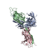 3dosC  3dpbC  1z9sS S: Starting model for refinement C: citing same article ( |
|---|---|
| Similar structure data |


-Links
 PDBj
PDBj
- Assembly
| Deposited unit | 
| |||||||||||||||||||||||||||||||||||||||||||||||||||||||||||||||||||||||||||||||||||||||||||||||||||||||||||||||||||||||||||||||||||||||||||||||||||||||||||||||||||||||||||||||||||||||||||||
|---|---|---|---|---|---|---|---|---|---|---|---|---|---|---|---|---|---|---|---|---|---|---|---|---|---|---|---|---|---|---|---|---|---|---|---|---|---|---|---|---|---|---|---|---|---|---|---|---|---|---|---|---|---|---|---|---|---|---|---|---|---|---|---|---|---|---|---|---|---|---|---|---|---|---|---|---|---|---|---|---|---|---|---|---|---|---|---|---|---|---|---|---|---|---|---|---|---|---|---|---|---|---|---|---|---|---|---|---|---|---|---|---|---|---|---|---|---|---|---|---|---|---|---|---|---|---|---|---|---|---|---|---|---|---|---|---|---|---|---|---|---|---|---|---|---|---|---|---|---|---|---|---|---|---|---|---|---|---|---|---|---|---|---|---|---|---|---|---|---|---|---|---|---|---|---|---|---|---|---|---|---|---|---|---|---|---|---|---|---|---|
| 1 | 
| |||||||||||||||||||||||||||||||||||||||||||||||||||||||||||||||||||||||||||||||||||||||||||||||||||||||||||||||||||||||||||||||||||||||||||||||||||||||||||||||||||||||||||||||||||||||||||||
| 2 | 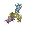
| |||||||||||||||||||||||||||||||||||||||||||||||||||||||||||||||||||||||||||||||||||||||||||||||||||||||||||||||||||||||||||||||||||||||||||||||||||||||||||||||||||||||||||||||||||||||||||||
| Unit cell |
| |||||||||||||||||||||||||||||||||||||||||||||||||||||||||||||||||||||||||||||||||||||||||||||||||||||||||||||||||||||||||||||||||||||||||||||||||||||||||||||||||||||||||||||||||||||||||||||
| Noncrystallographic symmetry (NCS) | NCS domain:
|