 Movie
Movie Controller

[English] 日本語
 Yorodumi
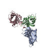
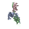
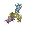
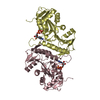
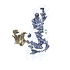
- PDB-3t72: PhoB(E)-Sigma70(4)-(RNAP-Betha-flap-tip-helix)-DNA Transcription ... -
+ Open data

- Basic information
| Entry | Database: PDB / ID: 3t72 | ||||||
|---|---|---|---|---|---|---|---|
| Title | PhoB(E)-Sigma70(4)-(RNAP-Betha-flap-tip-helix)-DNA Transcription Activation Sub-Complex | ||||||
 Components Components |
| ||||||
 Keywords Keywords | TRANSCRIPTION/DNA / WINGED-HELIX MOTIF / TRANSCRIPTION ACTIVATION / DNA-BINDING / TRANSCRIPTION-DNA complex | ||||||
| Function / homology |  Function and homology information Function and homology informationbacterial-type RNA polymerase holo enzyme binding / phosphate ion transport / sigma factor antagonist complex / phosphorelay response regulator activity / regulation of DNA-templated transcription initiation / sigma factor activity / cytosolic DNA-directed RNA polymerase complex / DNA-templated transcription initiation / protein-DNA complex / DNA-directed RNA polymerase activity ...bacterial-type RNA polymerase holo enzyme binding / phosphate ion transport / sigma factor antagonist complex / phosphorelay response regulator activity / regulation of DNA-templated transcription initiation / sigma factor activity / cytosolic DNA-directed RNA polymerase complex / DNA-templated transcription initiation / protein-DNA complex / DNA-directed RNA polymerase activity / response to heat / transcription cis-regulatory region binding / negative regulation of DNA-templated transcription / regulation of DNA-templated transcription / identical protein binding / cytosol Similarity search - Function | ||||||
| Biological species |  | ||||||
| Method |  X-RAY DIFFRACTION / SYNCHROTRON / MAD / Resolution: 4.33 Å X-RAY DIFFRACTION / SYNCHROTRON / MAD / Resolution: 4.33 Å | ||||||
 Authors Authors | Blanco, A.G. / Canals, A. / Bernues, J. / Sola, M. / Coll, M. | ||||||
 Citation Citation | Journal: Embo J. / Year: 2011 Title: The structure of a transcription activation subcomplex reveals how sigma (70) is recruited to PhoB promoters. Authors: Blanco, A.G. / Canals, A. / Bernues, J. / Sola, M. / Coll, M. | ||||||
| History |
|
- Structure visualization
| Structure viewer | Molecule: MolmilJmol/JSmol |
|---|
- Downloads & links
-Download
| PDBx/mmCIF format | 3t72.cif.gz | 407.7 KB | Display | PDBx/mmCIF format |
|---|---|---|---|---|
| PDB format | pdb3t72.ent.gz | 307.9 KB | Display | PDB format |
| PDBx/mmJSON format | 3t72.json.gz | Tree view | PDBx/mmJSON format | |
| Others |  Other downloads Other downloads |
-Validation report
| Arichive directory | https://data.pdbj.org/pub/pdb/validation_reports/t7/3t72ftp://data.pdbj.org/pub/pdb/validation_reports/t7/3t72 | HTTPS FTP |
|---|
-Related structure data
| Similar structure data |
|---|


-Links
 PDBj
PDBj


- Assembly
| Deposited unit | 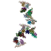
| ||||||||
|---|---|---|---|---|---|---|---|---|---|
| 1 | 
| ||||||||
| 2 | 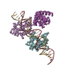
| ||||||||
| 3 | 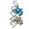
| ||||||||
| 4 | 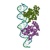
| ||||||||
| 5 | 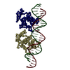
| ||||||||
| 6 | 
| ||||||||
| 7 | 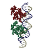
| ||||||||
| 8 | 
| ||||||||
| 9 | 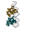
| ||||||||
| 10 | 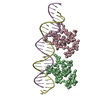
| ||||||||
| 11 | 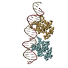
| ||||||||
| 12 | 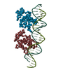
| ||||||||
| Unit cell |
| ||||||||
| Details | The biological assembly is that formed by chains A,B,C,D,q and r |
-Components
| #1: Protein | Mass: 11971.624 Da / Num. of mol.: 24 Source method: isolated from a genetically manipulated source Source: (gene. exp.) #2: DNA chain | Mass: 8035.216 Da / Num. of mol.: 12 / Source method: obtained synthetically / Details: Synthesized DNA / Source: (synth.) #3: DNA chain | Mass: 7937.140 Da / Num. of mol.: 12 / Source method: obtained synthetically / Details: Synthesized DNA / Source: (synth.) #4: Protein | Mass: 11080.619 Da / Num. of mol.: 2 Source method: isolated from a genetically manipulated source Source: (gene. exp.) References: UniProt: P00579, UniProt: E8Y6A0, DNA-directed RNA polymerase |
|---|
-Experimental details
-Experiment
| Experiment | Method: X-RAY DIFFRACTION / Number of used crystals: 1 |
|---|
- Sample preparation
| Crystal | Density Matthews: 5.81 Å3/Da / Density % sol: 78.81 % |
|---|---|
| Crystal grow | Temperature: 293 K / Method: vapor diffusion, sitting drop / pH: 6 Details: 8-10% PEG 4000, 100 mM KCl, 10 mM magnesium chloride, 50 mM MES, pH 6.0, VAPOR DIFFUSION, SITTING DROP, temperature 293K |
-Data collection
| Diffraction | Mean temperature: 200 K | ||||||||||||
|---|---|---|---|---|---|---|---|---|---|---|---|---|---|
| Diffraction source | Source: SYNCHROTRON / Site: ESRF  / Beamline: ID29 / Wavelength: 1.2542,1.2554,1.2498 / Beamline: ID29 / Wavelength: 1.2542,1.2554,1.2498 | ||||||||||||
| Detector | Type: MAR CCD 165 mm / Detector: CCD / Date: Jun 27, 2006 | ||||||||||||
| Radiation | Monochromator: GRAPHITE / Protocol: MAD / Monochromatic (M) / Laue (L): M / Scattering type: x-ray | ||||||||||||
| Radiation wavelength |
| ||||||||||||
| Reflection | Resolution: 4.33→50 Å / Num. all: 73615 / Num. obs: 73615 / % possible obs: 100 % / Observed criterion σ(F): 1.5 / Observed criterion σ(I): 1.5 |
- Processing
| Software |
| ||||||||||||
|---|---|---|---|---|---|---|---|---|---|---|---|---|---|
| Refinement | Method to determine structure: MAD / Resolution: 4.33→50 Å / Num. reflection obs: 73615 Details: CLOSE CONTACTS OF DNA ATOMS WITH SYMMETRY-EQUIVALENT NEIGHBOUR DNA MOLECULES FORMING PSEUDO-CONTINUOUS HELICES ARE DUE TO LACK OF ATOMIC POSITIONAL REFINEMENT | ||||||||||||
| Refinement step | Cycle: LAST / Resolution: 4.33→50 Å
|
