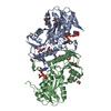
| Deposited unit | A: Ribose-phosphate pyrophosphokinase
B: Ribose-phosphate pyrophosphokinase
C: Ribose-phosphate pyrophosphokinase
D: Ribose-phosphate pyrophosphokinase
E: Ribose-phosphate pyrophosphokinase
F: Ribose-phosphate pyrophosphokinase
hetero molecules
| Theoretical mass | Number of molelcules |
|---|
| Total (without water) | 195,999 | 18 |
|---|
| Polymers | 193,339 | 6 |
|---|
| Non-polymers | 2,660 | 12 |
|---|
| Water | 432 | 24 |
|---|
|
|---|
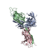
| 1 | A: Ribose-phosphate pyrophosphokinase
B: Ribose-phosphate pyrophosphokinase
hetero molecules
| Theoretical mass | Number of molelcules |
|---|
| Total (without water) | 65,333 | 6 |
|---|
| Polymers | 64,446 | 2 |
|---|
| Non-polymers | 887 | 4 |
|---|
| Water | 36 | 2 |
|---|
| Type | Name | Symmetry operation | Number |
|---|
| identity operation | 1_555 | x,y,z | 1 |
| Buried area | 5550 Å2 |
|---|
| ΔGint | -49 kcal/mol |
|---|
| Surface area | 22740 Å2 |
|---|
| Method | PISA |
|---|
|
|---|
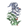
| 2 | C: Ribose-phosphate pyrophosphokinase
D: Ribose-phosphate pyrophosphokinase
hetero molecules
| Theoretical mass | Number of molelcules |
|---|
| Total (without water) | 65,333 | 6 |
|---|
| Polymers | 64,446 | 2 |
|---|
| Non-polymers | 887 | 4 |
|---|
| Water | 36 | 2 |
|---|
| Type | Name | Symmetry operation | Number |
|---|
| identity operation | 1_555 | x,y,z | 1 |
| Buried area | 5210 Å2 |
|---|
| ΔGint | -29 kcal/mol |
|---|
| Surface area | 22710 Å2 |
|---|
| Method | PISA |
|---|
|
|---|
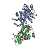
| 3 | E: Ribose-phosphate pyrophosphokinase
F: Ribose-phosphate pyrophosphokinase
hetero molecules
| Theoretical mass | Number of molelcules |
|---|
| Total (without water) | 65,333 | 6 |
|---|
| Polymers | 64,446 | 2 |
|---|
| Non-polymers | 887 | 4 |
|---|
| Water | 36 | 2 |
|---|
| Type | Name | Symmetry operation | Number |
|---|
| identity operation | 1_555 | x,y,z | 1 |
| Buried area | 5210 Å2 |
|---|
| ΔGint | -28 kcal/mol |
|---|
| Surface area | 22740 Å2 |
|---|
| Method | PISA |
|---|
|
|---|
| Unit cell | | Length a, b, c (Å) | 80.217, 92.846, 138.338 |
|---|
| Angle α, β, γ (deg.) | 90.00, 90.91, 90.00 |
|---|
| Int Tables number | 4 |
|---|
| Space group name H-M | P1211 |
|---|
|
|---|
| Noncrystallographic symmetry (NCS) | NCS domain: | ID | Ens-ID |
|---|
| 1 | 1 | | 2 | 1 | | 1 | 2 | | 2 | 2 | | 1 | 3 | | 2 | 3 | | 1 | 4 | | 2 | 4 | | 1 | 5 | | 2 | 5 |
NCS domain segments: | Dom-ID | Component-ID | Ens-ID | Selection details |
|---|
| 1 | 1 | 1 | chain A and (resseq 1:190 or resseq 204:291 or resseq 301:302 or resseq 402:405 )| 2 | 1 | 1 | chain B and (resseq 1:190 or resseq 204:291 or resseq 301:302 or resseq 402:405 )| 1 | 1 | 2 | chain A and (resseq 1:190 or resseq 204:291 or resseq 301:302 or resseq 402:405 )| 2 | 1 | 2 | chain C and (resseq 1:190 or resseq 204:291 or resseq 301:302 or resseq 402:405 )| 1 | 1 | 3 | chain A and (resseq 1:190 or resseq 204:291 or resseq 301:302 or resseq 402:405 )| 2 | 1 | 3 | chain D and (resseq 1:190 or resseq 204:291 or resseq 301:302 or resseq 402:405 )| 1 | 1 | 4 | chain A and (resseq 1:190 or resseq 204 | | | | | | |
|
|---|
 Movie
Movie Controller


 Open data
 Basic information
 Components
 Keywords
 Function and homology information
 Sulfolobus solfataricus (archaea)
 X-RAY DIFFRACTION /
  Authors
 Citation
 Structure visualization
 Downloads & links
 Other downloads


 PDBj
PDBj


 Assembly