 Movie
Movie Controller

[English] 日本語
 Yorodumi
- PDB-1nce: Crystal structure of a ternary complex of E. coli thymidylate syn... -
+ Open data

- Basic information
| Entry | Database: PDB / ID: 1nce | ||||||
|---|---|---|---|---|---|---|---|
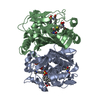
| Title | Crystal structure of a ternary complex of E. coli thymidylate synthase D169C with dUMP and the antifolate CB3717 | ||||||
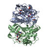
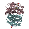
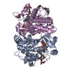
 Components Components | Thymidylate synthase | ||||||
 Keywords Keywords | BIOSYNTHETIC PROTEIN / beta-sheet interface / protein-dUMP-cofactor analog complex / asymmetric dimer | ||||||
| Function / homology |  Function and homology information Function and homology informationthymidylate synthase / thymidylate synthase activity / dTTP biosynthetic process / dTMP biosynthetic process / response to radiation / regulation of translation / methylation / magnesium ion binding / protein homodimerization activity / RNA binding / cytosol Similarity search - Function | ||||||
| Biological species |  | ||||||
| Method |  X-RAY DIFFRACTION / FOURIER SYNTHESIS / Resolution: 2.4 Å X-RAY DIFFRACTION / FOURIER SYNTHESIS / Resolution: 2.4 Å | ||||||
 Authors Authors | Birdsall, D.L. / Finer-Moore, J. / Stroud, R.M. | ||||||
 Citation Citation | Journal: Protein Eng. / Year: 2003 Title: The only active mutant of thymidylate synthase D169, a residue far from the site of methyl transfer, demonstrates the exquisite nature of enzyme specificity. Authors: Birdsall, D.L. / Finer-Moore, J. / Stroud, R.M. #1: Journal: Biochemistry / Year: 1998Title: D221 in thymidylate synthase controls conformation change, and thereby opening of the imidazolidine Authors: Sage, C.R. / Michelitsch, M.D. / Stout, T.J. / Biermann, D. / Nissen, R. / Finer-Moore, J. / Stroud, R.M. #2: Journal: Biochemistry / Year: 1990Title: Structure, multiple site binding, and segmental accommodation in thymidylate synthase on binding dUMP and an anti-folate Authors: Montfort, W.R. / Perry, K.M. / Fauman, E.B. / Finer-Moore, J.S. / Maley, G.F. / Hardy, L. / Maley, F. / Stroud, R.M. | ||||||
| History |
|
- Structure visualization
| Structure viewer | Molecule: MolmilJmol/JSmol |
|---|
- Downloads & links
-Download
| PDBx/mmCIF format | 1nce.cif.gz | 125 KB | Display | PDBx/mmCIF format |
|---|---|---|---|---|
| PDB format | pdb1nce.ent.gz | 97.4 KB | Display | PDB format |
| PDBx/mmJSON format | 1nce.json.gz | Tree view | PDBx/mmJSON format | |
| Others |  Other downloads Other downloads |
-Validation report
| Arichive directory | https://data.pdbj.org/pub/pdb/validation_reports/nc/1nceftp://data.pdbj.org/pub/pdb/validation_reports/nc/1nce | HTTPS FTP |
|---|
-Related structure data
| Related structure data | 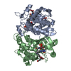 1kceS S: Starting model for refinement |
|---|---|
| Similar structure data |


-Links
 PDBj
PDBj- Assembly
| Deposited unit | 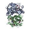
| ||||||||
|---|---|---|---|---|---|---|---|---|---|
| 1 |
| ||||||||
| Unit cell |
| ||||||||
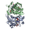
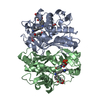
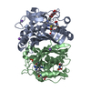
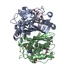
| Details | The biological assembly is a homodimer consisting of chains A and B |
-Components
| #1: Protein | Mass: 30547.721 Da / Num. of mol.: 2 / Mutation: D169C Source method: isolated from a genetically manipulated source Source: (gene. exp.) Escherichia coli, Escherichia coli O157:H7 Genus: Escherichia, Escherichia / Species: , Escherichia coli / Strain: , O157:H7 / Gene: THYA OR B2827 OR Z4144 OR ECS3684 / Production host: #2: Chemical |   Mass: 308.182 Da / Num. of mol.: 2 / Source method: obtained synthetically / Formula: C9H13N2O8P Mass: 308.182 Da / Num. of mol.: 2 / Source method: obtained synthetically / Formula: C9H13N2O8P#3: Chemical | 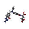  Mass: 477.469 Da / Num. of mol.: 2 / Source method: obtained synthetically / Formula: C24H23N5O6 Mass: 477.469 Da / Num. of mol.: 2 / Source method: obtained synthetically / Formula: C24H23N5O6#4: Water | ChemComp-HOH / |  Mass: 18.015 Da / Num. of mol.: 178 / Source method: isolated from a natural source / Formula: H2O Mass: 18.015 Da / Num. of mol.: 178 / Source method: isolated from a natural source / Formula: H2OHas protein modification | Y | |
|---|
-Experimental details
-Experiment
| Experiment | Method: X-RAY DIFFRACTION / Number of used crystals: 1 |
|---|
- Sample preparation
| Crystal | Density Matthews: 2.6 Å3/Da / Density % sol: 52.65 % | ||||||||||||||||||||||||||||||||||||||||||||||||||||||||||||
|---|---|---|---|---|---|---|---|---|---|---|---|---|---|---|---|---|---|---|---|---|---|---|---|---|---|---|---|---|---|---|---|---|---|---|---|---|---|---|---|---|---|---|---|---|---|---|---|---|---|---|---|---|---|---|---|---|---|---|---|---|---|
| Crystal grow | Temperature: 298 K / Method: vapor diffusion, hanging drop / pH: 9.1 Details: potassium phosphate, EDTA, ammonium sulfate, DTT, pH 9.1, VAPOR DIFFUSION, HANGING DROP, temperature 298.0K | ||||||||||||||||||||||||||||||||||||||||||||||||||||||||||||
| Crystal grow | *PLUS | ||||||||||||||||||||||||||||||||||||||||||||||||||||||||||||
| Components of the solutions | *PLUS
|
-Data collection
| Diffraction | Mean temperature: 298 K |
|---|---|
| Diffraction source | Source: ROTATING ANODE / Type: RIGAKU RU200 / Wavelength: 1.5418 |
| Detector | Type: RIGAKU RAXIS II / Detector: IMAGE PLATE / Date: Dec 21, 1998 / Details: monochromater |
| Radiation | Monochromator: graphite / Protocol: SINGLE WAVELENGTH / Monochromatic (M) / Laue (L): M / Scattering type: x-ray |
| Radiation wavelength | Wavelength: 1.5418 Å / Relative weight: 1 |
| Reflection | Resolution: 2.4→40 Å / Num. all: 24679 / Num. obs: 24679 / % possible obs: 100 % / Observed criterion σ(F): -3 / Observed criterion σ(I): -3 / Redundancy: 8.7 % / Rmerge(I) obs: 0.174 / Net I/σ(I): 8.1 |
| Reflection shell | Resolution: 2.4→2.55 Å / Mean I/σ(I) obs: 1.9 / % possible all: 100 |
| Reflection | *PLUS % possible obs: 100 % |
- Processing
| Software |
| ||||||||||||||||||||||||||||||||||||
|---|---|---|---|---|---|---|---|---|---|---|---|---|---|---|---|---|---|---|---|---|---|---|---|---|---|---|---|---|---|---|---|---|---|---|---|---|---|
| Refinement | Method to determine structure: FOURIER SYNTHESIS Starting model: PDB entry 1KCE with waters and ligands removed Resolution: 2.4→36.69 Å / Rfactor Rfree error: 0.006 / Isotropic thermal model: RESTRAINED / Cross valid method: THROUGHOUT / σ(F): 0 / σ(I): -3 / Stereochemistry target values: Engh & Huber
| ||||||||||||||||||||||||||||||||||||
| Solvent computation | Solvent model: FLAT MODEL / Bsol: 56.9659 Å2 / ksol: 0.334593 e/Å3 | ||||||||||||||||||||||||||||||||||||
| Displacement parameters | Biso mean: 34.2 Å2
| ||||||||||||||||||||||||||||||||||||
| Refine analyze | Luzzati coordinate error free: 0.37 Å / Luzzati sigma a free: 0.5 Å | ||||||||||||||||||||||||||||||||||||
| Refinement step | Cycle: LAST / Resolution: 2.4→36.69 Å
| ||||||||||||||||||||||||||||||||||||
| Refine LS restraints |
| ||||||||||||||||||||||||||||||||||||
| LS refinement shell | Resolution: 2.4→2.55 Å / Rfactor Rfree error: 0.02 / Total num. of bins used: 6
| ||||||||||||||||||||||||||||||||||||
| Xplor file |
| ||||||||||||||||||||||||||||||||||||
| Refinement | *PLUS Highest resolution: 2.4 Å | ||||||||||||||||||||||||||||||||||||
| Solvent computation | *PLUS | ||||||||||||||||||||||||||||||||||||
| Displacement parameters | *PLUS | ||||||||||||||||||||||||||||||||||||
| Refine LS restraints | *PLUS
| ||||||||||||||||||||||||||||||||||||
| LS refinement shell | *PLUS Highest resolution: 2.4 Å |
