 Movie
Movie Controller

[English] 日本語
 Yorodumi
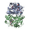
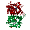
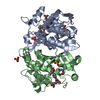
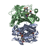
- PDB-1bjg: D221(169)N MUTANT DOES NOT PROMOTE OPENING OF THE COFACTOR IMIDAZ... -
+ Open data

- Basic information
| Entry | Database: PDB / ID: 1bjg | ||||||
|---|---|---|---|---|---|---|---|
| Title | D221(169)N MUTANT DOES NOT PROMOTE OPENING OF THE COFACTOR IMIDAZOLIDINE RING | ||||||
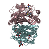
 Components Components | THYMIDYLATE SYNTHASE | ||||||
 Keywords Keywords | TRANSFERASE / ACTIVE SITE MUTANT / REACTION INTERMEDIATE METHYLTRANSFERASE | ||||||
| Function / homology |  Function and homology information Function and homology informationthymidylate synthase / thymidylate synthase activity / dTTP biosynthetic process / dTMP biosynthetic process / response to radiation / regulation of translation / methylation / magnesium ion binding / protein homodimerization activity / RNA binding / cytosol Similarity search - Function | ||||||
| Biological species |  | ||||||
| Method |  X-RAY DIFFRACTION / MOLECULAR REPLACEMENT / Resolution: 2.3 Å X-RAY DIFFRACTION / MOLECULAR REPLACEMENT / Resolution: 2.3 Å | ||||||
 Authors Authors | Sage, C.R. / Michelitsch, M.D. / Finer-Moore, J. / Stroud, R.M. | ||||||
 Citation Citation | Journal: Biochemistry / Year: 1998 Title: D221 in thymidylate synthase controls conformation change, and thereby opening of the imidazolidine. Authors: Sage, C.R. / Michelitsch, M.D. / Stout, T.J. / Biermann, D. / Nissen, R. / Finer-Moore, J. / Stroud, R.M. #1: Journal: Faseb J. / Year: 1993Title: Stereochemistry of a Multistep/Bipartite Methyl Transfer Reaction: Thymidylate Synthase Authors: Stroud, R.M. / Finer-Moore, J.S. #2: Journal: Biochemistry / Year: 1990Title: Structure, Multiple Site Binding, and Segmental Accommodation in Thymidylate Synthase on Binding Dump and an Anti-Folate Authors: Montfort, W.R. / Perry, K.M. / Fauman, E.B. / Finer-Moore, J.S. / Maley, G.F. / Hardy, L. / Maley, F. / Stroud, R.M. #3: Journal: Biochemistry / Year: 1990Title: Erratum. Structure, Multiple Site Binding, and Segmental Accommodation in Thymidylate Synthase on Binding Dump and an Anti-Folate Authors: Montfort, W.R. / Perry, K.M. / Fauman, E.B. / Finer-Moore, J.S. / Maley, G.F. / Hardy, L. / Maley, F. / Stroud, R.M. | ||||||
| History |
|
- Structure visualization
| Structure viewer | Molecule: MolmilJmol/JSmol |
|---|
- Downloads & links
-Download
| PDBx/mmCIF format | 1bjg.cif.gz | 71.6 KB | Display | PDBx/mmCIF format |
|---|---|---|---|---|
| PDB format | pdb1bjg.ent.gz | 53.4 KB | Display | PDB format |
| PDBx/mmJSON format | 1bjg.json.gz | Tree view | PDBx/mmJSON format | |
| Others |  Other downloads Other downloads |
-Validation report
| Arichive directory | https://data.pdbj.org/pub/pdb/validation_reports/bj/1bjgftp://data.pdbj.org/pub/pdb/validation_reports/bj/1bjg | HTTPS FTP |
|---|
-Related structure data


-Links
 PDBj
PDBj- Assembly
| Deposited unit | 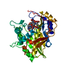
| |||||||||
|---|---|---|---|---|---|---|---|---|---|---|
| 1 | 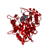
| |||||||||
| Unit cell |
| |||||||||
| Components on special symmetry positions |
|
-Components
| #1: Protein | Mass: 30558.678 Da / Num. of mol.: 1 / Mutation: D169N / Source method: isolated from a natural source / Source: (natural) |
|---|---|
| #2: Chemical | ChemComp-UFP / 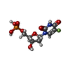  Type: DNA linking / Mass: 326.172 Da / Num. of mol.: 1 / Source method: obtained synthetically / Formula: C9H12FN2O8P / Comment: inhibitor*YM Type: DNA linking / Mass: 326.172 Da / Num. of mol.: 1 / Source method: obtained synthetically / Formula: C9H12FN2O8P / Comment: inhibitor*YM |
| #3: Chemical | ChemComp-TMF / 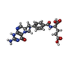  Mass: 455.424 Da / Num. of mol.: 1 / Source method: obtained synthetically / Formula: C20H21N7O6 Mass: 455.424 Da / Num. of mol.: 1 / Source method: obtained synthetically / Formula: C20H21N7O6 |
| #4: Water | ChemComp-HOH /  Mass: 18.015 Da / Num. of mol.: 92 / Source method: isolated from a natural source / Formula: H2O Mass: 18.015 Da / Num. of mol.: 92 / Source method: isolated from a natural source / Formula: H2O |
| Has protein modification | Y |
-Experimental details
-Experiment
| Experiment | Method: X-RAY DIFFRACTION / Number of used crystals: 1 |
|---|
- Sample preparation
| Crystal | Density Matthews: 3.21 Å3/Da / Density % sol: 60 % | ||||||||||||||||||||||||||||||||||||||||||
|---|---|---|---|---|---|---|---|---|---|---|---|---|---|---|---|---|---|---|---|---|---|---|---|---|---|---|---|---|---|---|---|---|---|---|---|---|---|---|---|---|---|---|---|
| Crystal grow | pH: 7.7 / Details: pH 7.7 | ||||||||||||||||||||||||||||||||||||||||||
| Crystal | *PLUS | ||||||||||||||||||||||||||||||||||||||||||
| Crystal grow | *PLUS Method: vapor diffusion / PH range low: 7.8 / PH range high: 7.6 | ||||||||||||||||||||||||||||||||||||||||||
| Components of the solutions | *PLUS
|
-Data collection
| Diffraction | Mean temperature: 294 K |
|---|---|
| Diffraction source | Source: ROTATING ANODE / Type: RIGAKU / Wavelength: 1.5418 |
| Detector | Type: RIGAKU / Detector: IMAGE PLATE / Date: Jan 1, 1997 |
| Radiation | Monochromator: GRAPHITE(002) / Monochromatic (M) / Laue (L): M / Scattering type: x-ray |
| Radiation wavelength | Wavelength: 1.5418 Å / Relative weight: 1 |
| Reflection | Highest resolution: 2.3 Å / Num. obs: 16757 / % possible obs: 94.9 % / Redundancy: 4 % / Biso Wilson estimate: 30.3 Å2 / Rmerge(I) obs: 0.075 / Rsym value: 0.075 / Net I/σ(I): 10.4 |
| Reflection shell | Resolution: 2.3→2.37 Å / Redundancy: 2.9 % / Rmerge(I) obs: 0.25 / Mean I/σ(I) obs: 3.6 / Rsym value: 0.25 / % possible all: 90.1 |
| Reflection | *PLUS Num. measured all: 68196 |
| Reflection shell | *PLUS % possible obs: 90.1 % |
- Processing
| Software |
| ||||||||||||||||||||||||||||||||||||||||||||||||||||||||||||||||||||||||||||||||
|---|---|---|---|---|---|---|---|---|---|---|---|---|---|---|---|---|---|---|---|---|---|---|---|---|---|---|---|---|---|---|---|---|---|---|---|---|---|---|---|---|---|---|---|---|---|---|---|---|---|---|---|---|---|---|---|---|---|---|---|---|---|---|---|---|---|---|---|---|---|---|---|---|---|---|---|---|---|---|---|---|---|
| Refinement | Method to determine structure: MOLECULAR REPLACEMENT Starting model: WILD TYPE E. COLI TS Resolution: 2.3→7 Å / Rfactor Rfree error: 0.006 / Data cutoff high absF: 100000000 / Data cutoff low absF: 0.001 / Isotropic thermal model: RESTRAINED / Cross valid method: THROUGHOUT / σ(F): 2
| ||||||||||||||||||||||||||||||||||||||||||||||||||||||||||||||||||||||||||||||||
| Displacement parameters | Biso mean: 26.3 Å2 | ||||||||||||||||||||||||||||||||||||||||||||||||||||||||||||||||||||||||||||||||
| Refine analyze |
| ||||||||||||||||||||||||||||||||||||||||||||||||||||||||||||||||||||||||||||||||
| Refinement step | Cycle: LAST / Resolution: 2.3→7 Å
| ||||||||||||||||||||||||||||||||||||||||||||||||||||||||||||||||||||||||||||||||
| Refine LS restraints |
| ||||||||||||||||||||||||||||||||||||||||||||||||||||||||||||||||||||||||||||||||
| LS refinement shell | Resolution: 2.3→2.44 Å / Rfactor Rfree error: 0.019 / Total num. of bins used: 6
| ||||||||||||||||||||||||||||||||||||||||||||||||||||||||||||||||||||||||||||||||
| Xplor file |
| ||||||||||||||||||||||||||||||||||||||||||||||||||||||||||||||||||||||||||||||||
| Software | *PLUS Name: X-PLOR / Version: 3.1 / Classification: refinement | ||||||||||||||||||||||||||||||||||||||||||||||||||||||||||||||||||||||||||||||||
| Refine LS restraints | *PLUS
|
