 Movie
Movie Controller


+ Open data

- Basic information
| Entry | Database: PDB / ID: 1kzj | ||||||
|---|---|---|---|---|---|---|---|
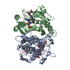
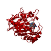
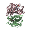
| Title | Crystal Structure of EcTS W80G/dUMP/CB3717 Complex | ||||||
 Components Components | Thymidylate synthase | ||||||
 Keywords Keywords | TRANSFERASE / Enzyme substrate cofactor analog complex | ||||||
| Function / homology |  Function and homology information Function and homology informationthymidylate synthase / thymidylate synthase activity / dTTP biosynthetic process / dTMP biosynthetic process / response to radiation / regulation of translation / methylation / magnesium ion binding / protein homodimerization activity / RNA binding / cytosol Similarity search - Function | ||||||
| Biological species |  | ||||||
| Method |  X-RAY DIFFRACTION / SYNCHROTRON / MOLECULAR REPLACEMENT / Resolution: 2.6 Å X-RAY DIFFRACTION / SYNCHROTRON / MOLECULAR REPLACEMENT / Resolution: 2.6 Å | ||||||
 Authors Authors | Fritz, T.A. / Liu, L. / Finer-Moore, J.S. / Stroud, R.M. | ||||||
 Citation Citation | Journal: Biochemistry / Year: 2002 Title: Tryptophan 80 and leucine 143 are critical for the hydride transfer step of thymidylate synthase by controlling active site access. Authors: Fritz, T.A. / Liu, L. / Finer-Moore, J.S. / Stroud, R.M. | ||||||
| History |
|
- Structure visualization
| Structure viewer | Molecule: MolmilJmol/JSmol |
|---|
- Downloads & links
-Download
| PDBx/mmCIF format | 1kzj.cif.gz | 331.3 KB | Display | PDBx/mmCIF format |
|---|---|---|---|---|
| PDB format | pdb1kzj.ent.gz | 272.2 KB | Display | PDB format |
| PDBx/mmJSON format | 1kzj.json.gz | Tree view | PDBx/mmJSON format | |
| Others |  Other downloads Other downloads |
-Validation report
| Arichive directory | https://data.pdbj.org/pub/pdb/validation_reports/kz/1kzjftp://data.pdbj.org/pub/pdb/validation_reports/kz/1kzj | HTTPS FTP |
|---|
-Related structure data
| Related structure data | 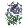 1kziC  2kceS C: citing same article ( S: Starting model for refinement |
|---|---|
| Similar structure data |


-Links
 PDBj
PDBj- Assembly
| Deposited unit | 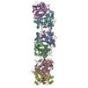
| ||||||||
|---|---|---|---|---|---|---|---|---|---|
| 1 | 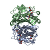
| ||||||||
| 2 | 
| ||||||||
| 3 | 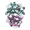
| ||||||||
| Unit cell |
| ||||||||
| Details | The biological unit consists of a dimer comprised of chains A and B or C and D or E and F of the asymmetric unit. |
-Components
| #1: Protein | Mass: 30430.506 Da / Num. of mol.: 6 / Mutation: W80G Source method: isolated from a genetically manipulated source Source: (gene. exp.) #2: Chemical | ChemComp-UMP /   Mass: 308.182 Da / Num. of mol.: 6 / Source method: obtained synthetically / Formula: C9H13N2O8P Mass: 308.182 Da / Num. of mol.: 6 / Source method: obtained synthetically / Formula: C9H13N2O8P#3: Chemical | ChemComp-CB3 /   Mass: 477.469 Da / Num. of mol.: 6 / Source method: obtained synthetically / Formula: C24H23N5O6 Mass: 477.469 Da / Num. of mol.: 6 / Source method: obtained synthetically / Formula: C24H23N5O6#4: Water | ChemComp-HOH / |  Mass: 18.015 Da / Num. of mol.: 292 / Source method: isolated from a natural source / Formula: H2O Mass: 18.015 Da / Num. of mol.: 292 / Source method: isolated from a natural source / Formula: H2OHas protein modification | Y | |
|---|
-Experimental details
-Experiment
| Experiment | Method: X-RAY DIFFRACTION / Number of used crystals: 1 |
|---|
- Sample preparation
| Crystal | Density Matthews: 2.3 Å3/Da / Density % sol: 46.45 % | ||||||||||||||||||||||||||||||||||||||||||||||||||||||
|---|---|---|---|---|---|---|---|---|---|---|---|---|---|---|---|---|---|---|---|---|---|---|---|---|---|---|---|---|---|---|---|---|---|---|---|---|---|---|---|---|---|---|---|---|---|---|---|---|---|---|---|---|---|---|---|
| Crystal grow | Temperature: 298 K / Method: vapor diffusion, hanging drop / pH: 8.5 Details: 0.1 M Tris, 22-26% PEG 4000, 0.2 M sodium acetate, 10 mM DTT, pH 8.5, VAPOR DIFFUSION, HANGING DROP, temperature 298K | ||||||||||||||||||||||||||||||||||||||||||||||||||||||
| Crystal grow | *PLUS | ||||||||||||||||||||||||||||||||||||||||||||||||||||||
| Components of the solutions | *PLUS
|
-Data collection
| Diffraction | Mean temperature: 100 K |
|---|---|
| Diffraction source | Source: SYNCHROTRON / Site: SSRL  / Beamline: BL7-1 / Wavelength: 1.08 Å / Beamline: BL7-1 / Wavelength: 1.08 Å |
| Detector | Type: MARRESEARCH / Detector: IMAGE PLATE / Date: Nov 20, 1999 |
| Radiation | Monochromator: Cylindrically bent, triangular, Si 111 asymmetric cut, horizontal focus Protocol: SINGLE WAVELENGTH / Monochromatic (M) / Laue (L): M / Scattering type: x-ray |
| Radiation wavelength | Wavelength: 1.08 Å / Relative weight: 1 |
| Reflection | Resolution: 2.6→25.35 Å / Num. obs: 51013 / % possible obs: 98.1 % / Observed criterion σ(I): -3 / Biso Wilson estimate: 26.3 Å2 |
| Reflection shell | Resolution: 2.6→2.69 Å / % possible all: 92.4 |
| Reflection | *PLUS Highest resolution: 2.6 Å / Num. measured all: 444846 / Rmerge(I) obs: 0.124 |
| Reflection shell | *PLUS % possible obs: 92.4 % / Rmerge(I) obs: 0.41 / Mean I/σ(I) obs: 2.4 |
- Processing
| Software |
| ||||||||||||||||||||
|---|---|---|---|---|---|---|---|---|---|---|---|---|---|---|---|---|---|---|---|---|---|
| Refinement | Method to determine structure: MOLECULAR REPLACEMENT Starting model: PDB entry 2KCE with ligands and water removed. Resolution: 2.6→25.35 Å / Rfactor Rfree error: 0.004 / Data cutoff high absF: 1552392.29 / Data cutoff low absF: 0 / Isotropic thermal model: RESTRAINED / Cross valid method: THROUGHOUT / σ(F): 0 / Stereochemistry target values: Engh & Huber
| ||||||||||||||||||||
| Solvent computation | Solvent model: FLAT MODEL / Bsol: 27.3867 Å2 / ksol: 0.350189 e/Å3 | ||||||||||||||||||||
| Displacement parameters | Biso mean: 20.8 Å2
| ||||||||||||||||||||
| Refine analyze |
| ||||||||||||||||||||
| Refinement step | Cycle: LAST / Resolution: 2.6→25.35 Å
| ||||||||||||||||||||
| Refine LS restraints |
| ||||||||||||||||||||
| LS refinement shell | Resolution: 2.6→2.76 Å / Rfactor Rfree error: 0.015 / Total num. of bins used: 6
| ||||||||||||||||||||
| Xplor file |
| ||||||||||||||||||||
| Refinement | *PLUS Highest resolution: 2.6 Å / Rfactor Rfree: 0.258 / Rfactor Rwork: 0.215 | ||||||||||||||||||||
| Solvent computation | *PLUS | ||||||||||||||||||||
| Displacement parameters | *PLUS | ||||||||||||||||||||
| Refine LS restraints | *PLUS
| ||||||||||||||||||||
| LS refinement shell | *PLUS Lowest resolution: 2.69 Å / Rfactor Rfree: 0.34 / Rfactor Rwork: 0.28 |
