 Movie
Movie Controller

[English] 日本語
 Yorodumi
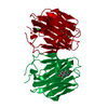
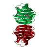
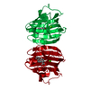
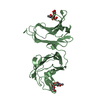
- PDB-1lhw: CRYSTAL STRUCTURE OF THE N-TERMINAL LG-DOMAIN OF SHBG IN COMPLEX ... -
+ Open data

- Basic information
| Entry | Database: PDB / ID: 1lhw | ||||||
|---|---|---|---|---|---|---|---|
| Title | CRYSTAL STRUCTURE OF THE N-TERMINAL LG-DOMAIN OF SHBG IN COMPLEX WITH 2-METHOXYESTRADIOL | ||||||
 Components Components | SEX HORMONE-BINDING GLOBULIN | ||||||
 Keywords Keywords | TRANSPORT PROTEIN / SHBG / 2-Methoxyestradiol | ||||||
| Function / homology |  Function and homology information Function and homology informationandrogen binding / steroid binding / extracellular exosome / extracellular region Similarity search - Function | ||||||
| Biological species |  Homo sapiens (human) Homo sapiens (human) | ||||||
| Method |  X-RAY DIFFRACTION / SYNCHROTRON / FOURIER SYNTHESIS / Resolution: 1.75 Å X-RAY DIFFRACTION / SYNCHROTRON / FOURIER SYNTHESIS / Resolution: 1.75 Å | ||||||
 Authors Authors | Avvakumov, G.V. / Grishkovskaya, I. / Muller, Y.A. / Hammond, G.L. | ||||||
 Citation Citation | Journal: J.Biol.Chem. / Year: 2002 Title: Crystal structure of human sex hormone-binding globulin in complex with 2-methoxyestradiol reveals the molecular basis for high affinity interactions with C-2 derivatives of estradiol. Authors: Avvakumov, G.V. / Grishkovskaya, I. / Muller, Y.A. / Hammond, G.L. | ||||||
| History |
|
- Structure visualization
| Structure viewer | Molecule: MolmilJmol/JSmol |
|---|
- Downloads & links
-Download
| PDBx/mmCIF format | 1lhw.cif.gz | 51.4 KB | Display | PDBx/mmCIF format |
|---|---|---|---|---|
| PDB format | pdb1lhw.ent.gz | 36.7 KB | Display | PDB format |
| PDBx/mmJSON format | 1lhw.json.gz | Tree view | PDBx/mmJSON format | |
| Others |  Other downloads Other downloads |
-Validation report
| Arichive directory | https://data.pdbj.org/pub/pdb/validation_reports/lh/1lhwftp://data.pdbj.org/pub/pdb/validation_reports/lh/1lhw | HTTPS FTP |
|---|
-Related structure data
| Related structure data |  1d2sS S: Starting model for refinement |
|---|---|
| Similar structure data |


-Links
 PDBj
PDBj

- Assembly
| Deposited unit | 
| ||||||||
|---|---|---|---|---|---|---|---|---|---|
| 1 | 
| ||||||||
| Unit cell |
|
-Components
| #1: Protein | Mass: 20972.832 Da / Num. of mol.: 1 / Fragment: LG-like 1 domain, Residues 30-218 Source method: isolated from a genetically manipulated source Source: (gene. exp.) Homo sapiens (human) / Plasmid: pGEX-2T / Production host:  |
|---|---|
| #2: Chemical | ChemComp-CA /   Mass: 40.078 Da / Num. of mol.: 1 / Source method: obtained synthetically / Formula: Ca Mass: 40.078 Da / Num. of mol.: 1 / Source method: obtained synthetically / Formula: Ca |
| #3: Chemical | ChemComp-ESM /   Mass: 302.408 Da / Num. of mol.: 1 / Source method: obtained synthetically / Formula: C19H26O3 Mass: 302.408 Da / Num. of mol.: 1 / Source method: obtained synthetically / Formula: C19H26O3 |
| #4: Chemical | ChemComp-IPA /   Mass: 60.095 Da / Num. of mol.: 1 / Source method: obtained synthetically / Formula: C3H8O / Comment: alkaloid*YM Mass: 60.095 Da / Num. of mol.: 1 / Source method: obtained synthetically / Formula: C3H8O / Comment: alkaloid*YM |
| #5: Water | ChemComp-HOH /  Mass: 18.015 Da / Num. of mol.: 97 / Source method: isolated from a natural source / Formula: H2O Mass: 18.015 Da / Num. of mol.: 97 / Source method: isolated from a natural source / Formula: H2O |
| Has protein modification | Y |
-Experimental details
-Experiment
| Experiment | Method: X-RAY DIFFRACTION / Number of used crystals: 1 |
|---|
- Sample preparation
| Crystal | Density Matthews: 2.09 Å3/Da / Density % sol: 41.17 % | |||||||||||||||||||||||||||||||||||||||||||||||||||||||||||||||
|---|---|---|---|---|---|---|---|---|---|---|---|---|---|---|---|---|---|---|---|---|---|---|---|---|---|---|---|---|---|---|---|---|---|---|---|---|---|---|---|---|---|---|---|---|---|---|---|---|---|---|---|---|---|---|---|---|---|---|---|---|---|---|---|---|
| Crystal grow | Temperature: 293 K / Method: vapor diffusion, hanging drop / pH: 7.5 Details: HEPES, Calcium chloride, 2-methoxyestradiol, pH 7.5, VAPOR DIFFUSION, HANGING DROP, temperature 293K | |||||||||||||||||||||||||||||||||||||||||||||||||||||||||||||||
| Crystal grow | *PLUS Temperature: 20 ℃ | |||||||||||||||||||||||||||||||||||||||||||||||||||||||||||||||
| Components of the solutions | *PLUS
|
-Data collection
| Diffraction | Mean temperature: 100 K |
|---|---|
| Diffraction source | Source: SYNCHROTRON / Site: EMBL/DESY, HAMBURG  / Beamline: BW7B / Wavelength: 0.8453 Å / Beamline: BW7B / Wavelength: 0.8453 Å |
| Detector | Type: MARRESEARCH / Detector: IMAGE PLATE / Date: May 5, 2001 |
| Radiation | Monochromator: triangular monochromator / Protocol: SINGLE WAVELENGTH / Monochromatic (M) / Laue (L): M / Scattering type: x-ray |
| Radiation wavelength | Wavelength: 0.8453 Å / Relative weight: 1 |
| Reflection | Resolution: 1.75→20 Å / Num. all: 17701 / Num. obs: 17701 / % possible obs: 99.4 % / Rmerge(I) obs: 0.031 |
| Reflection shell | Resolution: 1.75→1.79 Å / Rmerge(I) obs: 0.286 / % possible all: 96.9 |
| Reflection | *PLUS Lowest resolution: 20 Å / Num. measured all: 123818 |
| Reflection shell | *PLUS % possible obs: 96.9 % |
- Processing
| Software |
| ||||||||||||||||||||
|---|---|---|---|---|---|---|---|---|---|---|---|---|---|---|---|---|---|---|---|---|---|
| Refinement | Method to determine structure: FOURIER SYNTHESIS Starting model: PDB ENTRY 1D2S Resolution: 1.75→19.09 Å / Data cutoff high absF: 10000 / Data cutoff high rms absF: 10000 / Cross valid method: THROUGHOUT / σ(F): 0 / Stereochemistry target values: Engh & Huber
| ||||||||||||||||||||
| Refinement step | Cycle: LAST / Resolution: 1.75→19.09 Å
| ||||||||||||||||||||
| Refine LS restraints |
| ||||||||||||||||||||
| Refinement | *PLUS Lowest resolution: 20 Å / Rfactor Rwork: 0.21 | ||||||||||||||||||||
| Solvent computation | *PLUS | ||||||||||||||||||||
| Displacement parameters | *PLUS | ||||||||||||||||||||
| Refine LS restraints | *PLUS
|
