 Movie
Movie Controller


+ Open data

- Basic information
| Entry | Database: PDB / ID: 4grj | ||||||
|---|---|---|---|---|---|---|---|
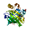
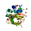
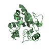
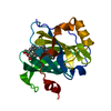
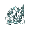
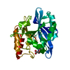
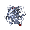
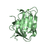
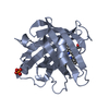
| Title | Crystal structure of nitrophorin 4 triple mutant complex with NO | ||||||
 Components Components | Nitrophorin-4 | ||||||
 Keywords Keywords | TRANSPORT PROTEIN / beta barrel / lipocalin / nitric oxide transport / nitric oxide binding | ||||||
| Function / homology |  Function and homology information Function and homology informationnitrite dismutase / histamine binding / nitric oxide binding / vasodilation / oxidoreductase activity / extracellular region / metal ion binding Similarity search - Function | ||||||
| Biological species |  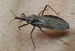 Rhodnius prolixus (insect) Rhodnius prolixus (insect) | ||||||
| Method |  X-RAY DIFFRACTION / SYNCHROTRON / FOURIER SYNTHESIS / Resolution: 1.15 Å X-RAY DIFFRACTION / SYNCHROTRON / FOURIER SYNTHESIS / Resolution: 1.15 Å | ||||||
 Authors Authors | Weichsel, A. / Issaian, A.V. / Montfort, W.R. | ||||||
 Citation Citation | Journal: To be Published Title: Crystal structures of nitrophorin 4 triple mutant Authors: Issaian, V.A. / Weichsel, A. / Montfort, W.R. #1: Journal: Structure / Year: 1998Title: The crystal structure of nitrophorin 4 at 1.5 resolution: transport of nitric oxide by a lipocalin-based heme protein Authors: Andersen, J.F. / Weichsel, A. / Balfour, C.A. / Champagne, D.E. / Montfort, W.R. #2: Journal: Biochemistry / Year: 2005Title: Ultrahigh resolution structures of nitrophorin 4: Heme distortion in ferrous CO and NO complexes Authors: Maes, E.M. / Roberts, S.A. / Weichsel, A. / Montfort, W.R. | ||||||
| History |
|
- Structure visualization
| Structure viewer | Molecule: MolmilJmol/JSmol |
|---|
- Downloads & links
-Download
| PDBx/mmCIF format | 4grj.cif.gz | 202.5 KB | Display | PDBx/mmCIF format |
|---|---|---|---|---|
| PDB format | pdb4grj.ent.gz | 165.1 KB | Display | PDB format |
| PDBx/mmJSON format | 4grj.json.gz | Tree view | PDBx/mmJSON format | |
| Others |  Other downloads Other downloads |
-Validation report
| Arichive directory | https://data.pdbj.org/pub/pdb/validation_reports/gr/4grjftp://data.pdbj.org/pub/pdb/validation_reports/gr/4grj | HTTPS FTP |
|---|
-Related structure data
| Related structure data |  4gnwSC S: Starting model for refinement C: citing same article ( |
|---|---|
| Similar structure data |


-Links
 PDBj
PDBj


- Assembly
| Deposited unit | 
| ||||||||
|---|---|---|---|---|---|---|---|---|---|
| 1 | 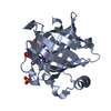
| ||||||||
| 2 | 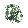
| ||||||||
| Unit cell |
|
-Components
| #1: Protein | Mass: 20289.709 Da / Num. of mol.: 2 / Mutation: D30N, E32Q, D35N Source method: isolated from a genetically manipulated source Source: (gene. exp.) Rhodnius prolixus (insect) / Plasmid: pET17b / Production host:  #2: Chemical |   Mass: 616.487 Da / Num. of mol.: 2 / Source method: obtained synthetically / Formula: C34H32FeN4O4 Mass: 616.487 Da / Num. of mol.: 2 / Source method: obtained synthetically / Formula: C34H32FeN4O4#3: Chemical |   Mass: 30.006 Da / Num. of mol.: 2 / Source method: obtained synthetically / Formula: NO Mass: 30.006 Da / Num. of mol.: 2 / Source method: obtained synthetically / Formula: NO#4: Chemical |   Mass: 94.971 Da / Num. of mol.: 2 / Source method: obtained synthetically / Formula: PO4 Mass: 94.971 Da / Num. of mol.: 2 / Source method: obtained synthetically / Formula: PO4#5: Water | ChemComp-HOH / |  Mass: 18.015 Da / Num. of mol.: 449 / Source method: isolated from a natural source / Formula: H2O Mass: 18.015 Da / Num. of mol.: 449 / Source method: isolated from a natural source / Formula: H2OHas protein modification | Y | |
|---|
-Experimental details
-Experiment
| Experiment | Method: X-RAY DIFFRACTION / Number of used crystals: 1 |
|---|
- Sample preparation
| Crystal | Density Matthews: 2.43 Å3/Da / Density % sol: 49.47 % |
|---|---|
| Crystal grow | Temperature: 298 K / Method: vapor diffusion, hanging drop / pH: 7.5 Details: 2.85 M ammonium phosphate, 100 mM Tris.HCl, pH 7.5, VAPOR DIFFUSION, HANGING DROP, temperature 298K |
-Data collection
| Diffraction | Mean temperature: 100 K |
|---|---|
| Diffraction source | Source: SYNCHROTRON / Site: SSRL  / Beamline: BL9-2 / Wavelength: 0.9795 Å / Beamline: BL9-2 / Wavelength: 0.9795 Å |
| Detector | Type: MARMOSAIC 325 mm CCD / Detector: CCD / Date: Jul 5, 2011 / Details: bent Si-mirror |
| Radiation | Monochromator: diamond (111) double-crystal / Protocol: SINGLE WAVELENGTH / Monochromatic (M) / Laue (L): M / Scattering type: x-ray |
| Radiation wavelength | Wavelength: 0.9795 Å / Relative weight: 1 |
| Reflection | Resolution: 1.15→60.31 Å / Num. all: 136529 / Num. obs: 136529 / % possible obs: 96.6 % / Observed criterion σ(F): 0 / Observed criterion σ(I): 0 / Redundancy: 4.9 % / Biso Wilson estimate: 19 Å2 / Rmerge(I) obs: 0.038 / Net I/σ(I): 12.1 |
| Reflection shell | Resolution: 1.15→1.19 Å / Redundancy: 4.8 % / Rmerge(I) obs: 0.54 / Mean I/σ(I) obs: 2.4 / Num. unique all: 13678 / % possible all: 98 |
- Processing
| Software |
| |||||||||||||||||||||||||||||||||||||||||||||||||||||||||||||||||||||||||||
|---|---|---|---|---|---|---|---|---|---|---|---|---|---|---|---|---|---|---|---|---|---|---|---|---|---|---|---|---|---|---|---|---|---|---|---|---|---|---|---|---|---|---|---|---|---|---|---|---|---|---|---|---|---|---|---|---|---|---|---|---|---|---|---|---|---|---|---|---|---|---|---|---|---|---|---|---|
| Refinement | Method to determine structure: FOURIER SYNTHESIS Starting model: PDB ENTRY 4GNW Resolution: 1.15→60.31 Å / Cor.coef. Fo:Fc: 0.977 / Cor.coef. Fo:Fc free: 0.973 / SU B: 1.342 / SU ML: 0.027 / Isotropic thermal model: anisotropic / Cross valid method: THROUGHOUT / σ(F): 0 / ESU R: 0.039 / ESU R Free: 0.038 / Stereochemistry target values: MAXIMUM LIKELIHOOD / Details: HYDROGENS HAVE BEEN ADDED IN THE RIDING POSITIONS
| |||||||||||||||||||||||||||||||||||||||||||||||||||||||||||||||||||||||||||
| Solvent computation | Ion probe radii: 0.8 Å / Shrinkage radii: 0.8 Å / VDW probe radii: 1.2 Å / Solvent model: MASK | |||||||||||||||||||||||||||||||||||||||||||||||||||||||||||||||||||||||||||
| Displacement parameters | Biso mean: 16.535 Å2
| |||||||||||||||||||||||||||||||||||||||||||||||||||||||||||||||||||||||||||
| Refine analyze | Luzzati coordinate error obs: 0.177 Å | |||||||||||||||||||||||||||||||||||||||||||||||||||||||||||||||||||||||||||
| Refinement step | Cycle: LAST / Resolution: 1.15→60.31 Å
| |||||||||||||||||||||||||||||||||||||||||||||||||||||||||||||||||||||||||||
| Refine LS restraints |
| |||||||||||||||||||||||||||||||||||||||||||||||||||||||||||||||||||||||||||
| LS refinement shell | Resolution: 1.15→1.18 Å / Total num. of bins used: 20
|
