 Movie
Movie Controller

[English] 日本語
 Yorodumi
- PDB-3u5r: Crystal structure of a hypothetical protein SMc02350 from Sinorhi... -
+ Open data

- Basic information
| Entry | Database: PDB / ID: 3u5r | ||||||
|---|---|---|---|---|---|---|---|
| Title | Crystal structure of a hypothetical protein SMc02350 from Sinorhizobium meliloti 1021 | ||||||
 Components Components | uncharacterized protein | ||||||
 Keywords Keywords | Structural Genomics / Unknown Function / PSI-Biology / New York Structural Genomics Research Consortium / NYSGRC / hypothetical protein / SMc02350 | ||||||
| Function / homology |  Function and homology information Function and homology information | ||||||
| Biological species |  Sinorhizobium meliloti (bacteria) Sinorhizobium meliloti (bacteria) | ||||||
| Method |  X-RAY DIFFRACTION / SYNCHROTRON / MOLECULAR REPLACEMENT / Resolution: 2.05 Å X-RAY DIFFRACTION / SYNCHROTRON / MOLECULAR REPLACEMENT / Resolution: 2.05 Å | ||||||
 Authors Authors | Agarwal, R. / Almo, S.C. / Swaminathan, S. / New York Structural Genomics Research Consortium (NYSGRC) | ||||||
 Citation Citation | Journal: To be Published Title: Crystal structure of a hypothetical protein SMc02350 from Sinorhizobium meliloti 1021 Authors: Agarwal, R. / Almo, S.C. / Swaminathan, S. | ||||||
| History |
|
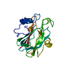
- Structure visualization
| Structure viewer | Molecule: MolmilJmol/JSmol |
|---|
- Downloads & links
-Download
| PDBx/mmCIF format | 3u5r.cif.gz | 156.4 KB | Display | PDBx/mmCIF format |
|---|---|---|---|---|
| PDB format | pdb3u5r.ent.gz | 123.1 KB | Display | PDB format |
| PDBx/mmJSON format | 3u5r.json.gz | Tree view | PDBx/mmJSON format | |
| Others |  Other downloads Other downloads |
-Validation report
| Arichive directory | https://data.pdbj.org/pub/pdb/validation_reports/u5/3u5rftp://data.pdbj.org/pub/pdb/validation_reports/u5/3u5r | HTTPS FTP |
|---|
-Related structure data
| Related structure data |  2ywiS S: Starting model for refinement |
|---|---|
| Similar structure data | |
| Other databases |


-Links
 PDBj
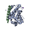
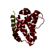
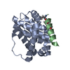
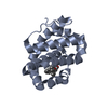
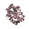
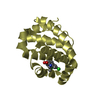
PDBj- Assembly
| Deposited unit | 
| ||||||||
|---|---|---|---|---|---|---|---|---|---|
| 1 | 
| ||||||||
| 2 | 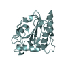
| ||||||||
| 3 | 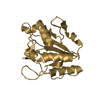
| ||||||||
| 4 | 
| ||||||||
| Unit cell |
|
-Components
| #1: Protein | Mass: 23515.324 Da / Num. of mol.: 4 Source method: isolated from a genetically manipulated source Source: (gene. exp.) Sinorhizobium meliloti (bacteria) / Strain: 1021 / Gene: R02577, SMc02350 / Plasmid: pET / Production host: #2: Water | ChemComp-HOH / |  Mass: 18.015 Da / Num. of mol.: 395 / Source method: isolated from a natural source / Formula: H2O Mass: 18.015 Da / Num. of mol.: 395 / Source method: isolated from a natural source / Formula: H2O |
|---|
-Experimental details
-Experiment
| Experiment | Method: X-RAY DIFFRACTION / Number of used crystals: 1 |
|---|
- Sample preparation
| Crystal | Density Matthews: 2.68 Å3/Da / Density % sol: 54.17 % |
|---|---|
| Crystal grow | Temperature: 293 K / Method: vapor diffusion, sitting drop / pH: 5.6 Details: 0.2M Ammonium acetate, 0.1M tri sodium citrate pH 5.6, 30% PEG 4000, VAPOR DIFFUSION, SITTING DROP, temperature 293K |
-Data collection
| Diffraction | Mean temperature: 100 K |
|---|---|
| Diffraction source | Source: SYNCHROTRON / Site: NSLS  / Beamline: X29A / Wavelength: 0.9792 Å / Beamline: X29A / Wavelength: 0.9792 Å |
| Detector | Type: ADSC QUANTUM 315 / Detector: CCD / Date: Oct 10, 2011 / Details: mirrors |
| Radiation | Monochromator: SI-III / Protocol: SINGLE WAVELENGTH / Monochromatic (M) / Laue (L): M / Scattering type: x-ray |
| Radiation wavelength | Wavelength: 0.9792 Å / Relative weight: 1 |
| Reflection | Resolution: 2.05→50 Å / Num. all: 59768 / Num. obs: 59768 / % possible obs: 97.9 % / Observed criterion σ(I): 0 / Redundancy: 4.4 % / Rmerge(I) obs: 0.05 / Net I/σ(I): 13.2 |
| Reflection shell | Resolution: 2.05→2.12 Å / Redundancy: 4.3 % / Rmerge(I) obs: 0.27 / Mean I/σ(I) obs: 5 / Num. unique all: 5911 / % possible all: 96.5 |
- Processing
| Software |
| |||||||||||||||||||||||||||||||||||||||||||||||||||||||||||||||||
|---|---|---|---|---|---|---|---|---|---|---|---|---|---|---|---|---|---|---|---|---|---|---|---|---|---|---|---|---|---|---|---|---|---|---|---|---|---|---|---|---|---|---|---|---|---|---|---|---|---|---|---|---|---|---|---|---|---|---|---|---|---|---|---|---|---|---|
| Refinement | Method to determine structure: MOLECULAR REPLACEMENT Starting model: pdb entry 2YWI Resolution: 2.05→34.02 Å / Cor.coef. Fo:Fc: 0.952 / Cor.coef. Fo:Fc free: 0.925 / SU B: 3.724 / SU ML: 0.103 / Cross valid method: THROUGHOUT / σ(I): 0 / ESU R Free: 0.159 / Stereochemistry target values: MAXIMUM LIKELIHOOD / Details: HYDROGENS HAVE BEEN ADDED IN THE RIDING POSITIONS
| |||||||||||||||||||||||||||||||||||||||||||||||||||||||||||||||||
| Solvent computation | Ion probe radii: 0.8 Å / Shrinkage radii: 0.8 Å / VDW probe radii: 1.4 Å / Solvent model: MASK | |||||||||||||||||||||||||||||||||||||||||||||||||||||||||||||||||
| Displacement parameters | Biso mean: 26.28 Å2
| |||||||||||||||||||||||||||||||||||||||||||||||||||||||||||||||||
| Refinement step | Cycle: LAST / Resolution: 2.05→34.02 Å
| |||||||||||||||||||||||||||||||||||||||||||||||||||||||||||||||||
| Refine LS restraints |
| |||||||||||||||||||||||||||||||||||||||||||||||||||||||||||||||||
| LS refinement shell | Resolution: 2.053→2.106 Å / Total num. of bins used: 20
|
