 Movie
Movie Controller

[English] 日本語
 Yorodumi
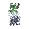
- PDB-1f5f: CRYSTAL STRUCTURE OF THE N-TERMINAL G-DOMAIN OF SHBG IN COMPLEX W... -
+ Open data

- Basic information
| Entry | Database: PDB / ID: 1f5f | ||||||
|---|---|---|---|---|---|---|---|
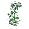
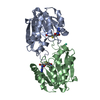
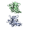
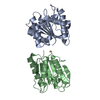
| Title | CRYSTAL STRUCTURE OF THE N-TERMINAL G-DOMAIN OF SHBG IN COMPLEX WITH ZINC | ||||||
 Components Components | SEX HORMONE-BINDING GLOBULIN | ||||||
 Keywords Keywords | SIGNALING PROTEIN / Jellyroll | ||||||
| Function / homology |  Function and homology information Function and homology informationandrogen binding / steroid binding / extracellular exosome / extracellular region Similarity search - Function | ||||||
| Biological species |  Homo sapiens (human) Homo sapiens (human) | ||||||
| Method |  X-RAY DIFFRACTION / SYNCHROTRON / Resolution: 1.7 Å X-RAY DIFFRACTION / SYNCHROTRON / Resolution: 1.7 Å | ||||||
 Authors Authors | Avvakumov, V.A. / Muller, Y.A. / Hammond, G.L. | ||||||
 Citation Citation | Journal: J.Biol.Chem. / Year: 2000 Title: Steroid-binding specificity of human sex hormone-binding globulin is influenced by occupancy of a zinc-binding site. Authors: Avvakumov, G.V. / Muller, Y.A. / Hammond, G.L. #1: Journal: Embo J. / Year: 2000Title: Crystal Structure of Human Sex Hormone-Binding Globulin: Steroid Transport by a Laminin G-Like Domain Authors: Grishkovskaya, I. / Avvakumov, G.V. / Sklenar, G. / Dales, D. / Hammond, G.L. / Muller, Y.A. | ||||||
| History |
|
- Structure visualization
| Structure viewer | Molecule: MolmilJmol/JSmol |
|---|
- Downloads & links
-Download
| PDBx/mmCIF format | 1f5f.cif.gz | 53.1 KB | Display | PDBx/mmCIF format |
|---|---|---|---|---|
| PDB format | pdb1f5f.ent.gz | 37.7 KB | Display | PDB format |
| PDBx/mmJSON format | 1f5f.json.gz | Tree view | PDBx/mmJSON format | |
| Others |  Other downloads Other downloads |
-Validation report
| Arichive directory | https://data.pdbj.org/pub/pdb/validation_reports/f5/1f5fftp://data.pdbj.org/pub/pdb/validation_reports/f5/1f5f | HTTPS FTP |
|---|
-Related structure data
| Related structure data | |
|---|---|
| Similar structure data |


-Links
 PDBj
PDBj

- Assembly
| Deposited unit | 
| ||||||||
|---|---|---|---|---|---|---|---|---|---|
| 1 | 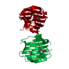
| ||||||||
| Unit cell |
| ||||||||
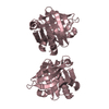
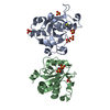
| Details | The biological assembly is a dimer constructed from chain A and a symmetry partner generated by a two-fold. |
-Components
-Protein , 1 types, 1 molecules A
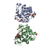
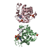
| #1: Protein | Mass: 22610.623 Da / Num. of mol.: 1 / Fragment: N-TERMINAL FRAGMENT OF SHBG (RESIDUES 1 TO 205) Source method: isolated from a genetically manipulated source Source: (gene. exp.) Homo sapiens (human) / Plasmid: PGEX-2T / Production host:  |
|---|
-Non-polymers , 5 types, 135 molecules 


| #2: Chemical | ChemComp-CA /  Mass: 40.078 Da / Num. of mol.: 1 / Source method: obtained synthetically / Formula: Ca Mass: 40.078 Da / Num. of mol.: 1 / Source method: obtained synthetically / Formula: Ca | ||||||
|---|---|---|---|---|---|---|---|
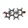
| #3: Chemical |  Mass: 65.409 Da / Num. of mol.: 2 / Source method: obtained synthetically / Formula: Zn Mass: 65.409 Da / Num. of mol.: 2 / Source method: obtained synthetically / Formula: Zn#4: Chemical | ChemComp-DHT / |  Mass: 290.440 Da / Num. of mol.: 1 / Source method: obtained synthetically / Formula: C19H30O2 / Comment: hormone*YM Mass: 290.440 Da / Num. of mol.: 1 / Source method: obtained synthetically / Formula: C19H30O2 / Comment: hormone*YM#5: Chemical | ChemComp-IPA / |  Mass: 60.095 Da / Num. of mol.: 1 / Source method: obtained synthetically / Formula: C3H8O Mass: 60.095 Da / Num. of mol.: 1 / Source method: obtained synthetically / Formula: C3H8O#6: Water | ChemComp-HOH / | Mass: 18.015 Da / Num. of mol.: 130 / Source method: isolated from a natural source / Formula: H2O |
-Details
| Has protein modification | Y |
|---|
-Experimental details
-Experiment
| Experiment | Method: X-RAY DIFFRACTION / Number of used crystals: 1 |
|---|
- Sample preparation
| Crystal | Density Matthews: 1.95 Å3/Da / Density % sol: 36.88 % | ||||||||||||||||||||||||||||||||||||||||||||||||
|---|---|---|---|---|---|---|---|---|---|---|---|---|---|---|---|---|---|---|---|---|---|---|---|---|---|---|---|---|---|---|---|---|---|---|---|---|---|---|---|---|---|---|---|---|---|---|---|---|---|
| Crystal grow | Temperature: 293.15 K / Method: vapor diffusion, hanging drop / pH: 7.5 Details: Crystal growth: ISOPROPANOL, PEG400, HEPES-buffer, CaCl2, DHT Crystals were soaked with 2.5 mM ZnCl2 for three days, pH 7.5, VAPOR DIFFUSION, HANGING DROP, temperature 293.15K | ||||||||||||||||||||||||||||||||||||||||||||||||
| Crystal grow | *PLUS Temperature: 293 KDetails: Grishkovskaya, I., (1999) Acta Crystallogr. Sec., D55, 2053 | ||||||||||||||||||||||||||||||||||||||||||||||||
| Components of the solutions | *PLUS
|
-Data collection
| Diffraction | Mean temperature: 100 K |
|---|---|
| Diffraction source | Source: SYNCHROTRON / Site: ESRF  / Type: ESRF / Wavelength: 0.933 / Type: ESRF / Wavelength: 0.933 |
| Detector | Type: MARRESEARCH / Detector: CCD / Date: Dec 8, 1999 |
| Radiation | Protocol: SINGLE WAVELENGTH / Monochromatic (M) / Laue (L): M / Scattering type: x-ray |
| Radiation wavelength | Wavelength: 0.933 Å / Relative weight: 1 |
| Reflection | Resolution: 1.7→20 Å / Num. all: 18785 / Num. obs: 18785 / % possible obs: 96.3 % / Observed criterion σ(F): 0 / Observed criterion σ(I): 0 / Redundancy: 5.4 % / Biso Wilson estimate: 35.8 Å2 / Rmerge(I) obs: 0.065 / Net I/σ(I): 13.9 |
| Reflection shell | Resolution: 1.7→1.8 Å / Redundancy: 3.7 % / Rmerge(I) obs: 0.287 / Num. unique all: 2825 / % possible all: 90.7 |
| Reflection | *PLUS Num. measured all: 101085 |
| Reflection shell | *PLUS % possible obs: 93.3 % / Mean I/σ(I) obs: 2.8 |
- Processing
| Software |
| ||||||||||||||||||||||||||||
|---|---|---|---|---|---|---|---|---|---|---|---|---|---|---|---|---|---|---|---|---|---|---|---|---|---|---|---|---|---|
| Refinement | Resolution: 1.7→20 Å / σ(F): 0 / σ(I): 0 / Stereochemistry target values: Engh & Huber
| ||||||||||||||||||||||||||||
| Displacement parameters | Biso mean: 38.7 Å2 | ||||||||||||||||||||||||||||
| Refinement step | Cycle: LAST / Resolution: 1.7→20 Å
| ||||||||||||||||||||||||||||
| Refine LS restraints |
| ||||||||||||||||||||||||||||
| Software | *PLUS Name: REFMAC / Classification: refinement | ||||||||||||||||||||||||||||
| Refinement | *PLUS Highest resolution: 1.7 Å / Lowest resolution: 20 Å / σ(F): 0 / Rfactor obs: 0.197 | ||||||||||||||||||||||||||||
| Solvent computation | *PLUS | ||||||||||||||||||||||||||||
| Displacement parameters | *PLUS | ||||||||||||||||||||||||||||
| Refine LS restraints | *PLUS
| ||||||||||||||||||||||||||||
| LS refinement shell | *PLUS Rfactor Rfree: 0.353 / Rfactor obs: 0.285 |
