 Movie
Movie Controller

[English] 日本語
 Yorodumi
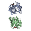
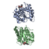
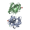
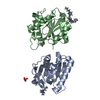
- PDB-1i6w: THE CRYSTAL STRUCTURE OF BACILLUS SUBTILIS LIPASE: A MINIMAL ALPH... -
+ Open data

- Basic information
| Entry | Database: PDB / ID: 1i6w | ||||||
|---|---|---|---|---|---|---|---|
| Title | THE CRYSTAL STRUCTURE OF BACILLUS SUBTILIS LIPASE: A MINIMAL ALPHA/BETA HYDROLASE ENZYME | ||||||
 Components Components | LIPASE A | ||||||
 Keywords Keywords | HYDROLASE / alpha/beta hydrolase | ||||||
| Function / homology |  Function and homology information Function and homology informationlipase activity / triacylglycerol lipase / triacylglycerol lipase activity / lipid catabolic process / extracellular region Similarity search - Function | ||||||
| Biological species |  | ||||||
| Method |  X-RAY DIFFRACTION / SYNCHROTRON / MIR / Resolution: 1.5 Å X-RAY DIFFRACTION / SYNCHROTRON / MIR / Resolution: 1.5 Å | ||||||
 Authors Authors | van Pouderoyen, G. / Eggert, T. / Jaeger, K.-E. / Dijkstra, B.W. | ||||||
 Citation Citation | Journal: J.Mol.Biol. / Year: 2001 Title: The crystal structure of Bacillus subtilis lipase: a minimal alpha/beta hydrolase fold enzyme. Authors: van Pouderoyen, G. / Eggert, T. / Jaeger, K.E. / Dijkstra, B.W. | ||||||
| History |
|
- Structure visualization
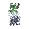
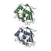
| Structure viewer | Molecule: MolmilJmol/JSmol |
|---|
- Downloads & links
-Download
| PDBx/mmCIF format | 1i6w.cif.gz | 89.6 KB | Display | PDBx/mmCIF format |
|---|---|---|---|---|
| PDB format | pdb1i6w.ent.gz | 67.6 KB | Display | PDB format |
| PDBx/mmJSON format | 1i6w.json.gz | Tree view | PDBx/mmJSON format | |
| Others |  Other downloads Other downloads |
-Validation report
| Arichive directory | https://data.pdbj.org/pub/pdb/validation_reports/i6/1i6wftp://data.pdbj.org/pub/pdb/validation_reports/i6/1i6w | HTTPS FTP |
|---|
-Related structure data
| Similar structure data |
|---|

-Links
 PDBj
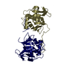
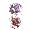
PDBj- Assembly
| Deposited unit | 
| ||||||||
|---|---|---|---|---|---|---|---|---|---|
| 1 |
| ||||||||
| Unit cell |
|
-Components
| #1: Protein | Mass: 19382.836 Da / Num. of mol.: 2 Source method: isolated from a genetically manipulated source Source: (gene. exp.) #2: Chemical | ChemComp-CD / |   Mass: 112.411 Da / Num. of mol.: 1 / Source method: obtained synthetically / Formula: Cd Mass: 112.411 Da / Num. of mol.: 1 / Source method: obtained synthetically / Formula: Cd#3: Water | ChemComp-HOH / |  Mass: 18.015 Da / Num. of mol.: 517 / Source method: isolated from a natural source / Formula: H2O Mass: 18.015 Da / Num. of mol.: 517 / Source method: isolated from a natural source / Formula: H2O |
|---|
-Experimental details
-Experiment
| Experiment | Method: X-RAY DIFFRACTION / Number of used crystals: 1 |
|---|
- Sample preparation
| Crystal | Density Matthews: 2.02 Å3/Da / Density % sol: 39 % | ||||||||||||||||||||||||||||||||||||||||||
|---|---|---|---|---|---|---|---|---|---|---|---|---|---|---|---|---|---|---|---|---|---|---|---|---|---|---|---|---|---|---|---|---|---|---|---|---|---|---|---|---|---|---|---|
| Crystal grow | Temperature: 293 K / Method: vapor diffusion, hanging drop / pH: 10 Details: PEG 4000, ethanol amine, sodium sulphate, cadmium chloride, pH 10.0, VAPOR DIFFUSION, HANGING DROP, temperature 293K | ||||||||||||||||||||||||||||||||||||||||||
| Crystal grow | *PLUS pH: 10 / Method: vapor diffusion | ||||||||||||||||||||||||||||||||||||||||||
| Components of the solutions | *PLUS
|
-Data collection
| Diffraction | Mean temperature: 100 K |
|---|---|
| Diffraction source | Source: SYNCHROTRON / Site: ESRF  / Beamline: ID14-4 / Wavelength: 0.9393 Å / Beamline: ID14-4 / Wavelength: 0.9393 Å |
| Detector | Type: ADSC QUANTUM 4 / Detector: CCD / Date: Dec 9, 1999 / Details: mirrors |
| Radiation | Monochromator: Si111 or Si311 crystals, LN2 cooled / Protocol: SINGLE WAVELENGTH / Monochromatic (M) / Laue (L): M / Scattering type: x-ray |
| Radiation wavelength | Wavelength: 0.9393 Å / Relative weight: 1 |
| Reflection | Resolution: 1.5→40 Å / Num. all: 41851 / Num. obs: 689356 / % possible obs: 82.4 % / Observed criterion σ(F): 0 / Observed criterion σ(I): 0 / Redundancy: 16.5 % / Biso Wilson estimate: 13.39 Å2 / Rsym value: 7.6 / Net I/σ(I): 20.8 |
| Reflection shell | Resolution: 1.5→1.53 Å / Redundancy: 2 % / Mean I/σ(I) obs: 2.7 / Num. unique all: 765 / Rsym value: 28 / % possible all: 30.6 |
| Reflection | *PLUS Num. obs: 41851 / Num. measured all: 689356 / Rmerge(I) obs: 0.076 |
| Reflection shell | *PLUS % possible obs: 30.6 % / Rmerge(I) obs: 0.28 |
- Processing
| Software |
| |||||||||||||||||||||||||
|---|---|---|---|---|---|---|---|---|---|---|---|---|---|---|---|---|---|---|---|---|---|---|---|---|---|---|
| Refinement | Method to determine structure: MIR Starting model: none Resolution: 1.5→40 Å / Isotropic thermal model: isotropic / Cross valid method: R-free / σ(F): 0 / σ(I): 0 / Stereochemistry target values: CNS_TOPPAR:protein_rep.param
| |||||||||||||||||||||||||
| Solvent computation | Solvent model: CNS ANISOTROPIC | |||||||||||||||||||||||||
| Displacement parameters | Biso mean: 15.4 Å2 | |||||||||||||||||||||||||
| Refine analyze |
| |||||||||||||||||||||||||
| Refinement step | Cycle: LAST / Resolution: 1.5→40 Å
| |||||||||||||||||||||||||
| Refine LS restraints |
| |||||||||||||||||||||||||
| LS refinement shell | Resolution: 1.5→1.51 Å / Total num. of bins used: 41
| |||||||||||||||||||||||||
| Software | *PLUS Name: CNS / Classification: refinement | |||||||||||||||||||||||||
| Refinement | *PLUS Highest resolution: 1.5 Å / Lowest resolution: 40 Å / σ(F): 0 / % reflection Rfree: 5 % | |||||||||||||||||||||||||
| Solvent computation | *PLUS | |||||||||||||||||||||||||
| Displacement parameters | *PLUS Biso mean: 15.4 Å2 | |||||||||||||||||||||||||
| Refine LS restraints | *PLUS
| |||||||||||||||||||||||||
| LS refinement shell | *PLUS % reflection Rfree: 5 % |
