 Movie
Movie Controller

[English] 日本語
 Yorodumi
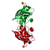
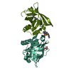
- PDB-2akq: The structure of bovine B-lactoglobulin A in crystals grown at ve... -
+ Open data

- Basic information
| Entry | Database: PDB / ID: 2akq | |||||||||
|---|---|---|---|---|---|---|---|---|---|---|
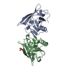
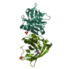
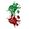
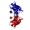
| Title | The structure of bovine B-lactoglobulin A in crystals grown at very low ionic strength | |||||||||
 Components Components | Beta-lactoglobulin variant A | |||||||||
 Keywords Keywords | TRANSPORT PROTEIN / B-lactoglbulin / crystal lattice / low ionic strength / pseudo-merohedral twinning | |||||||||
| Function / homology |  Function and homology information Function and homology informationretinol binding / long-chain fatty acid binding / extracellular region / identical protein binding Similarity search - Function | |||||||||
| Biological species |  | |||||||||
| Method |  X-RAY DIFFRACTION / MOLECULAR REPLACEMENT / Resolution: 3 Å X-RAY DIFFRACTION / MOLECULAR REPLACEMENT / Resolution: 3 Å | |||||||||
 Authors Authors | Adams, J.J. / Anderson, B.F. / Norris, G.E. / Creamer, L.K. / Jameson, G.B. | |||||||||
 Citation Citation | Journal: J.Struct.Biol. / Year: 2006 Title: Structure of bovine beta-lactoglobulin (variant A) at very low ionic strength Authors: Adams, J.J. / Anderson, B.F. / Norris, G.E. / Creamer, L.K. / Jameson, G.B. #1: Journal: Structure / Year: 1997Title: Bovine beta-lactoglobulin at 1.8 A resolution--still an enigmatic lipocalin Authors: Brownlow, S. / Morais-Cabral, J.H. / Cooper, R. / Flower, D.R. / Yewdall, S.J. / Polikarpov, I. / North, A.C. / Sawyer, L. #2: Journal: Biochemistry / Year: 1998Title: Structural basis of the Tanford transition of bovine beta-lactoglobulin Authors: Qin, B.Y. / Bewley, M.C. / Creamer, L.K. / Baker, H.M. / Baker, E.N. / Jameson, G.B. #3: Journal: Febs Lett. / Year: 1998Title: 12-Bromododecanoic acid binds inside the calyx of bovine beta-lactoglobulin Authors: Qin, B.Y. / Creamer, L.K. / Baker, E.N. / Jameson, G.B. | |||||||||
| History |
|
- Structure visualization
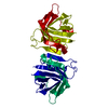
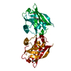
| Structure viewer | Molecule: MolmilJmol/JSmol |
|---|
- Downloads & links
-Download
| PDBx/mmCIF format | 2akq.cif.gz | 130 KB | Display | PDBx/mmCIF format |
|---|---|---|---|---|
| PDB format | pdb2akq.ent.gz | 103.6 KB | Display | PDB format |
| PDBx/mmJSON format | 2akq.json.gz | Tree view | PDBx/mmJSON format | |
| Others |  Other downloads Other downloads |
-Validation report
| Arichive directory | https://data.pdbj.org/pub/pdb/validation_reports/ak/2akqftp://data.pdbj.org/pub/pdb/validation_reports/ak/2akq | HTTPS FTP |
|---|
-Related structure data
| Related structure data |  1bebS S: Starting model for refinement |
|---|---|
| Similar structure data |


-Links
 PDBj
PDBj

- Assembly
| Deposited unit | 
| ||||||||
|---|---|---|---|---|---|---|---|---|---|
| 1 | 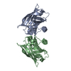
| ||||||||
| 2 | 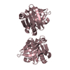
| ||||||||
| 3 | 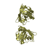
| ||||||||
| Unit cell |
|
-Components
| #1: Protein | Mass: 18387.264 Da / Num. of mol.: 4 / Source method: isolated from a natural source / Source: (natural) #2: Water | ChemComp-HOH / |  Mass: 18.015 Da / Num. of mol.: 68 / Source method: isolated from a natural source / Formula: H2O Mass: 18.015 Da / Num. of mol.: 68 / Source method: isolated from a natural source / Formula: H2OHas protein modification | Y | Sequence details | IN THE SWISS-PROT ENTRY, THE 64TH RESIDUE IS GLY, THE 118TH IS ALA. IN VARIANT A, THE 64TH IS ASP ...IN THE SWISS-PROT ENTRY, THE 64TH RESIDUE IS GLY, THE 118TH IS ALA. IN VARIANT A, THE 64TH IS ASP AND THE 118TH IS VAL. | |
|---|
-Experimental details
-Experiment
| Experiment | Method: X-RAY DIFFRACTION / Number of used crystals: 3 |
|---|
- Sample preparation
| Crystal | Density Matthews: 2.1 Å3/Da / Density % sol: 40.3 % |
|---|---|
| Crystal grow | Temperature: 295 K / Method: microdialysis / pH: 5.2 Details: Ultra pure water, pH 5.20, MICRODIALYSIS, temperature 295K |
-Data collection
| Diffraction | Mean temperature: 115 K |
|---|---|
| Diffraction source | Source: ROTATING ANODE / Type: RIGAKU RU200 / Wavelength: 1.5418 / Wavelength: 1.5418 Å |
| Detector | Type: RIGAKU RAXIS IIC / Detector: IMAGE PLATE / Date: Feb 15, 2001 |
| Radiation | Monochromator: AXCO P50 FOCUSSING CAPILLARY / Protocol: SINGLE WAVELENGTH / Monochromatic (M) / Laue (L): M / Scattering type: x-ray |
| Radiation wavelength | Wavelength: 1.5418 Å / Relative weight: 1 |
| Reflection | Resolution: 3→50 Å / Num. all: 12501 / Num. obs: 12501 / % possible obs: 92.6 % / Observed criterion σ(F): 4 / Observed criterion σ(I): 2 / Redundancy: 12 % / Rmerge(I) obs: 0.097 / Net I/σ(I): 10.3 |
| Reflection shell | Resolution: 3→3.11 Å / Redundancy: 3 % / Rmerge(I) obs: 0.298 / Mean I/σ(I) obs: 3 / Num. unique all: 1162 / % possible all: 94.3 |
- Processing
| Software |
| |||||||||||||||||||||||||||||||||
|---|---|---|---|---|---|---|---|---|---|---|---|---|---|---|---|---|---|---|---|---|---|---|---|---|---|---|---|---|---|---|---|---|---|---|
| Refinement | Method to determine structure: MOLECULAR REPLACEMENT Starting model: 1BEB Resolution: 3→10 Å / Num. parameters: 19369 / Num. restraintsaints: 35963 / Cross valid method: FREE R / σ(F): 0 / Stereochemistry target values: ENGH AND HUBER Details: This entry is a revision of 1mfh, taking into account 2 the effects of twinning. The twin law is (0 1 0/1 0 0/0 0 -1) R 3 is 0.325. Essentially addition of one parameter for twinning 4 ...Details: This entry is a revision of 1mfh, taking into account 2 the effects of twinning. The twin law is (0 1 0/1 0 0/0 0 -1) R 3 is 0.325. Essentially addition of one parameter for twinning 4 reduced R(working) from 0.293 to 0.224 and R(free) from 0.316 5 to 0.266. About torsion angles outside the expected ramachandran regions, TYR 99 is the central residue of a gamma turn characteristic of beta-lactoglobulins. ALA 34 is part of the dimer interface.
| |||||||||||||||||||||||||||||||||
| Refine analyze | Num. disordered residues: 7 / Occupancy sum hydrogen: 0 / Occupancy sum non hydrogen: 5017.5 | |||||||||||||||||||||||||||||||||
| Refinement step | Cycle: LAST / Resolution: 3→10 Å
| |||||||||||||||||||||||||||||||||
| Refine LS restraints |
| |||||||||||||||||||||||||||||||||
| LS refinement shell | Resolution: 3→3 Å
|
