 Movie
Movie Controller

[English] 日本語
 Yorodumi
- PDB-7kp5: Energetic and structural effects of the Tanford transition on the... -
+ Open data

- Basic information
| Entry | Database: PDB / ID: 7kp5 | ||||||
|---|---|---|---|---|---|---|---|
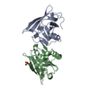
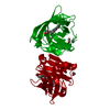
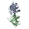
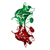
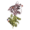
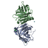
| Title | Energetic and structural effects of the Tanford transition on the ligand recognition of bovine Beta-lactoglobulin | ||||||
 Components Components | Beta-lactoglobulin | ||||||
 Keywords Keywords | TRANSPORT PROTEIN / Bovine beta-lactoglobulin / Lipocalin / Tanford transition / Structural energetics | ||||||
| Function / homology |  Function and homology information Function and homology informationretinol binding / long-chain fatty acid binding / extracellular region / identical protein binding Similarity search - Function | ||||||
| Biological species |  | ||||||
| Method |  X-RAY DIFFRACTION / MOLECULAR REPLACEMENT / Resolution: 2.4 Å X-RAY DIFFRACTION / MOLECULAR REPLACEMENT / Resolution: 2.4 Å | ||||||
 Authors Authors | Rodriguez-Hernandez, A. / Rodriguez-Romero, A. | ||||||
| Funding support |  Mexico, 1items Mexico, 1items
| ||||||
 Citation Citation | Journal: Arch.Biochem.Biophys. / Year: 2021 Title: Energetic and structural effects of the Tanford transition on ligand recognition of bovine beta-lactoglobulin. Authors: Labra-Nunez, A. / Cofas-Vargas, L.F. / Gutierrez-Magdaleno, G. / Gomez-Velasco, H. / Rodriguez-Hernandez, A. / Rodriguez-Romero, A. / Garcia-Hernandez, E. | ||||||
| History |
|
- Structure visualization
| Structure viewer | Molecule: MolmilJmol/JSmol |
|---|
- Downloads & links
-Download
| PDBx/mmCIF format | 7kp5.cif.gz | 89 KB | Display | PDBx/mmCIF format |
|---|---|---|---|---|
| PDB format | pdb7kp5.ent.gz | Display | PDB format | |
| PDBx/mmJSON format | 7kp5.json.gz | Tree view | PDBx/mmJSON format | |
| Others |  Other downloads Other downloads |
-Validation report
| Arichive directory | https://data.pdbj.org/pub/pdb/validation_reports/kp/7kp5ftp://data.pdbj.org/pub/pdb/validation_reports/kp/7kp5 | HTTPS FTP |
|---|
-Related structure data
| Related structure data | 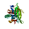 7kotC  1bebS C: citing same article ( S: Starting model for refinement |
|---|---|
| Similar structure data |


-Links
 PDBj
PDBj

- Assembly
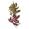
| Deposited unit | 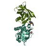
| ||||||||||||
|---|---|---|---|---|---|---|---|---|---|---|---|---|---|
| 1 |
| ||||||||||||
| Unit cell |
|
-Components
| #1: Protein | Mass: 18301.174 Da / Num. of mol.: 2 / Source method: isolated from a natural source / Source: (natural) #2: Chemical |   Mass: 266.397 Da / Num. of mol.: 3 / Source method: obtained synthetically / Formula: C12H26O4S / Feature type: SUBJECT OF INVESTIGATION / Comment: detergent*YM Mass: 266.397 Da / Num. of mol.: 3 / Source method: obtained synthetically / Formula: C12H26O4S / Feature type: SUBJECT OF INVESTIGATION / Comment: detergent*YM#3: Water | ChemComp-HOH / |  Mass: 18.015 Da / Num. of mol.: 35 / Source method: isolated from a natural source / Formula: H2O Mass: 18.015 Da / Num. of mol.: 35 / Source method: isolated from a natural source / Formula: H2OHas ligand of interest | Y | Has protein modification | Y | |
|---|
-Experimental details
-Experiment
| Experiment | Method: X-RAY DIFFRACTION / Number of used crystals: 1 |
|---|
- Sample preparation
| Crystal | Density Matthews: 2.39 Å3/Da / Density % sol: 48.56 % |
|---|---|
| Crystal grow | Temperature: 277.15 K / Method: vapor diffusion, hanging drop / pH: 4.5 Details: 12-15 mg/mL protein in 0.05M acetate and 0.1 M NaCl. Combined 1:1 with 0.05M KH2PO4, 20% PEG |
-Data collection
| Diffraction | Mean temperature: 100 K / Serial crystal experiment: N |
|---|---|
| Diffraction source | Source: ROTATING ANODE / Type: RIGAKU MICROMAX-007 HF / Wavelength: 1.54 Å |
| Detector | Type: DECTRIS PILATUS 200K / Detector: PIXEL / Date: Jul 3, 2017 |
| Radiation | Protocol: SINGLE WAVELENGTH / Monochromatic (M) / Laue (L): M / Scattering type: x-ray |
| Radiation wavelength | Wavelength: 1.54 Å / Relative weight: 1 |
| Reflection | Resolution: 2.39→38.86 Å / Num. obs: 14623 / % possible obs: 99.9 % / Redundancy: 7.3 % / Biso Wilson estimate: 49.5 Å2 / CC1/2: 0.95 / Net I/σ(I): 35.7 |
| Reflection shell | Resolution: 2.39→2.43 Å / Num. unique obs: 697 / CC1/2: 0.85 |
- Processing
| Software |
| ||||||||||||||||||||||||||||||||||||||||||||||||||||||||
|---|---|---|---|---|---|---|---|---|---|---|---|---|---|---|---|---|---|---|---|---|---|---|---|---|---|---|---|---|---|---|---|---|---|---|---|---|---|---|---|---|---|---|---|---|---|---|---|---|---|---|---|---|---|---|---|---|---|
| Refinement | Method to determine structure: MOLECULAR REPLACEMENT Starting model: 1BEB Resolution: 2.4→38.86 Å / SU ML: 0.2816 / Cross valid method: FREE R-VALUE / σ(F): 1.34 / Phase error: 27.6508 Stereochemistry target values: GeoStd + Monomer Library + CDL v1.2
| ||||||||||||||||||||||||||||||||||||||||||||||||||||||||
| Solvent computation | Shrinkage radii: 0.9 Å / VDW probe radii: 1.11 Å / Solvent model: FLAT BULK SOLVENT MODEL | ||||||||||||||||||||||||||||||||||||||||||||||||||||||||
| Displacement parameters | Biso mean: 54.76 Å2 | ||||||||||||||||||||||||||||||||||||||||||||||||||||||||
| Refinement step | Cycle: LAST / Resolution: 2.4→38.86 Å
| ||||||||||||||||||||||||||||||||||||||||||||||||||||||||
| Refine LS restraints |
| ||||||||||||||||||||||||||||||||||||||||||||||||||||||||
| LS refinement shell |
|
