 Movie
Movie Controller

[English] 日本語
 Yorodumi
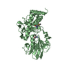
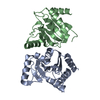
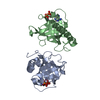
- PDB-2liv: PERIPLASMIC BINDING PROTEIN STRUCTURE AND FUNCTION. REFINED X-RAY... -
+ Open data

- Basic information
| Entry | Database: PDB / ID: 2liv | ||||||
|---|---|---|---|---|---|---|---|
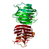
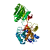
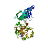
| Title | PERIPLASMIC BINDING PROTEIN STRUCTURE AND FUNCTION. REFINED X-RAY STRUCTURES OF THE LEUCINE/ISOLEUCINE/VALINE-BINDING PROTEIN AND ITS COMPLEX WITH LEUCINE | ||||||
 Components Components | LEUCINE | ||||||
 Keywords Keywords | PERIPLASMIC BINDING PROTEIN | ||||||
| Function / homology |  Function and homology information Function and homology informationbranched-chain amino acid transport / valine transport / isoleucine transport / L-leucine transport / amino acid transport / ATP-binding cassette (ABC) transporter complex, substrate-binding subunit-containing / outer membrane-bounded periplasmic space / periplasmic space / membrane Similarity search - Function | ||||||
| Biological species |  | ||||||
| Method |  X-RAY DIFFRACTION / Resolution: 2.4 Å X-RAY DIFFRACTION / Resolution: 2.4 Å | ||||||
 Authors Authors | Sack, J.S. / Saper, M.A. / Quiocho, F.A. | ||||||
 Citation Citation | Journal: J.Mol.Biol. / Year: 1989 Title: Periplasmic binding protein structure and function. Refined X-ray structures of the leucine/isoleucine/valine-binding protein and its complex with leucine. Authors: Sack, J.S. / Saper, M.A. / Quiocho, F.A. #1: Journal: J.Mol.Biol. / Year: 1989Title: Structure of the L-Leucine-Binding Protein Refined at 2.4 Angstroms Resolution and Comparison with the Leu(Slash)Ile(Slash)Val-Binding Protein Structure Authors: Sack, J.S. / Trakhanov, S.D. / Tsigannik, I.H. / Quiocho, F.A. #2: Journal: J.Biol.Chem. / Year: 1983Title: Leucine, Isoleucine, Valine-Binding Protein from Escherichia Coli Authors: Saper, M.A. / Quiocho, F.A. | ||||||
| History |
|
- Structure visualization
| Structure viewer | Molecule: MolmilJmol/JSmol |
|---|
- Downloads & links
-Download
| PDBx/mmCIF format | 2liv.cif.gz | 78.2 KB | Display | PDBx/mmCIF format |
|---|---|---|---|---|
| PDB format | pdb2liv.ent.gz | 58.3 KB | Display | PDB format |
| PDBx/mmJSON format | 2liv.json.gz | Tree view | PDBx/mmJSON format | |
| Others |  Other downloads Other downloads |
-Validation report
| Arichive directory | https://data.pdbj.org/pub/pdb/validation_reports/li/2livftp://data.pdbj.org/pub/pdb/validation_reports/li/2liv | HTTPS FTP |
|---|
-Related structure data
| Similar structure data |
|---|


-Links
 PDBj
PDBj- Assembly
| Deposited unit | 
| ||||||||
|---|---|---|---|---|---|---|---|---|---|
| 1 |
| ||||||||
| Unit cell |
|
-Components
| #1: Protein | Mass: 36784.344 Da / Num. of mol.: 1 Source method: isolated from a genetically manipulated source Source: (gene. exp.) |
|---|---|
| #2: Water | ChemComp-HOH /  Mass: 18.015 Da / Num. of mol.: 121 / Source method: isolated from a natural source / Formula: H2O Mass: 18.015 Da / Num. of mol.: 121 / Source method: isolated from a natural source / Formula: H2O |
| Has protein modification | Y |
-Experimental details
-Experiment
| Experiment | Method: X-RAY DIFFRACTION |
|---|
- Sample preparation
| Crystal | Density Matthews: 2.22 Å3/Da / Density % sol: 44.69 % | ||||||||||||||||||||||||||||||||||||||||||||||||||||||||||||
|---|---|---|---|---|---|---|---|---|---|---|---|---|---|---|---|---|---|---|---|---|---|---|---|---|---|---|---|---|---|---|---|---|---|---|---|---|---|---|---|---|---|---|---|---|---|---|---|---|---|---|---|---|---|---|---|---|---|---|---|---|---|
| Crystal grow | *PLUS pH: 7.6 / Method: other | ||||||||||||||||||||||||||||||||||||||||||||||||||||||||||||
| Components of the solutions | *PLUS
|
-Data collection
| Reflection | *PLUS Highest resolution: 2.4 Å / Num. obs: 12004 / Num. measured all: 41179 / Rmerge(I) obs: 0.021 |
|---|
- Processing
| Software | Name: PROLSQ / Classification: refinement | ||||||||||||||||||||||||||||||||||||||||||||||||||||||||||||||||||||||||||||||||||||
|---|---|---|---|---|---|---|---|---|---|---|---|---|---|---|---|---|---|---|---|---|---|---|---|---|---|---|---|---|---|---|---|---|---|---|---|---|---|---|---|---|---|---|---|---|---|---|---|---|---|---|---|---|---|---|---|---|---|---|---|---|---|---|---|---|---|---|---|---|---|---|---|---|---|---|---|---|---|---|---|---|---|---|---|---|---|
| Refinement | Resolution: 2.4→10 Å /
| ||||||||||||||||||||||||||||||||||||||||||||||||||||||||||||||||||||||||||||||||||||
| Refinement step | Cycle: LAST / Resolution: 2.4→10 Å
| ||||||||||||||||||||||||||||||||||||||||||||||||||||||||||||||||||||||||||||||||||||
| Refine LS restraints |
| ||||||||||||||||||||||||||||||||||||||||||||||||||||||||||||||||||||||||||||||||||||
| Refinement | *PLUS Highest resolution: 2.4 Å / Lowest resolution: 10 Å / Num. reflection all: 11817 / Rfactor all: 0.179 | ||||||||||||||||||||||||||||||||||||||||||||||||||||||||||||||||||||||||||||||||||||
| Solvent computation | *PLUS | ||||||||||||||||||||||||||||||||||||||||||||||||||||||||||||||||||||||||||||||||||||
| Displacement parameters | *PLUS |
