 Movie
Movie Controller

[English] 日本語
 Yorodumi
- PDB-1kdk: THE STRUCTURE OF THE N-TERMINAL LG DOMAIN OF SHBG IN CRYSTALS SOA... -
+ Open data

- Basic information
| Entry | Database: PDB / ID: 1kdk | ||||||
|---|---|---|---|---|---|---|---|
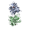
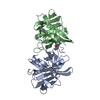
| Title | THE STRUCTURE OF THE N-TERMINAL LG DOMAIN OF SHBG IN CRYSTALS SOAKED WITH EDTA | ||||||
 Components Components | Sex Hormone-Binding Globulin | ||||||
 Keywords Keywords | TRANSPORT PROTEIN / SHBG / DHT | ||||||
| Function / homology |  Function and homology information Function and homology informationandrogen binding / steroid binding / extracellular exosome / extracellular region Similarity search - Function | ||||||
| Biological species |  Homo sapiens (human) Homo sapiens (human) | ||||||
| Method |  X-RAY DIFFRACTION / SYNCHROTRON / FOURIER SYNTHESIS / Resolution: 1.7 Å X-RAY DIFFRACTION / SYNCHROTRON / FOURIER SYNTHESIS / Resolution: 1.7 Å | ||||||
 Authors Authors | Grishkovskaya, I. / Avvakumov, G.V. / Hammond, G.L. / Muller, Y.A. | ||||||
 Citation Citation | Journal: J.Mol.Biol. / Year: 2002 Title: Resolution of a disordered region at the entrance of the human sex hormone-binding globulin steroid-binding site. Authors: Grishkovskaya, I. / Avvakumov, G.V. / Hammond, G.L. / Muller, Y.A. | ||||||
| History |
|
- Structure visualization
| Structure viewer | Molecule: MolmilJmol/JSmol |
|---|
- Downloads & links
-Download
| PDBx/mmCIF format | 1kdk.cif.gz | 50.8 KB | Display | PDBx/mmCIF format |
|---|---|---|---|---|
| PDB format | pdb1kdk.ent.gz | 35.9 KB | Display | PDB format |
| PDBx/mmJSON format | 1kdk.json.gz | Tree view | PDBx/mmJSON format | |
| Others |  Other downloads Other downloads |
-Validation report
| Arichive directory | https://data.pdbj.org/pub/pdb/validation_reports/kd/1kdkftp://data.pdbj.org/pub/pdb/validation_reports/kd/1kdk | HTTPS FTP |
|---|
-Related structure data
| Related structure data | 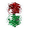 1kdmC  1d2sS S: Starting model for refinement C: citing same article ( |
|---|---|
| Similar structure data |


-Links
 PDBj
PDBj
- Assembly
| Deposited unit | 
| ||||||||
|---|---|---|---|---|---|---|---|---|---|
| 1 | 
| ||||||||
| Unit cell |
|
-Components
| #1: Protein | Mass: 19655.336 Da / Num. of mol.: 1 / Fragment: N-terminal LG-domain Source method: isolated from a genetically manipulated source Source: (gene. exp.) Homo sapiens (human) / Plasmid: pGEX-2T / Production host:  |
|---|---|
| #2: Chemical | ChemComp-DHT /   Mass: 290.440 Da / Num. of mol.: 1 / Source method: obtained synthetically / Formula: C19H30O2 Mass: 290.440 Da / Num. of mol.: 1 / Source method: obtained synthetically / Formula: C19H30O2 |
| #3: Water | ChemComp-HOH /  Mass: 18.015 Da / Num. of mol.: 89 / Source method: isolated from a natural source / Formula: H2O Mass: 18.015 Da / Num. of mol.: 89 / Source method: isolated from a natural source / Formula: H2O |
| Has protein modification | Y |
-Experimental details
-Experiment
| Experiment | Method: X-RAY DIFFRACTION / Number of used crystals: 1 |
|---|
- Sample preparation
| Crystal | Density Matthews: 2.26 Å3/Da / Density % sol: 45.5 % | ||||||||||||||||||||||||||||||||||||||||||||||||||||||||
|---|---|---|---|---|---|---|---|---|---|---|---|---|---|---|---|---|---|---|---|---|---|---|---|---|---|---|---|---|---|---|---|---|---|---|---|---|---|---|---|---|---|---|---|---|---|---|---|---|---|---|---|---|---|---|---|---|---|
| Crystal grow | Temperature: 293 K / Method: vapor diffusion, hanging drop / pH: 7.5 Details: PEG400, Isopropanol, pH 7.5, VAPOR DIFFUSION, HANGING DROP, temperature 293K | ||||||||||||||||||||||||||||||||||||||||||||||||||||||||
| Crystal grow | *PLUS Details: Grishkovskaya, I., (1999) Acta Crystallog. D55, 2053. | ||||||||||||||||||||||||||||||||||||||||||||||||||||||||
| Components of the solutions | *PLUS
|
-Data collection
| Diffraction | Mean temperature: 100 K |
|---|---|
| Diffraction source | Source: SYNCHROTRON / Site: ESRF  / Beamline: BM14 / Wavelength: 0.93 Å / Beamline: BM14 / Wavelength: 0.93 Å |
| Detector | Type: MARRESEARCH / Detector: IMAGE PLATE |
| Radiation | Monochromator: diamond(111), Ge(220) / Protocol: SINGLE WAVELENGTH / Monochromatic (M) / Laue (L): M / Scattering type: x-ray |
| Radiation wavelength | Wavelength: 0.93 Å / Relative weight: 1 |
| Reflection | Resolution: 1.7→40 Å / Num. all: 19262 / Num. obs: 19262 / % possible obs: 98.1 % / Observed criterion σ(F): 0 / Observed criterion σ(I): 0 / Redundancy: 4.7 % / Biso Wilson estimate: 33.1 Å2 / Rmerge(I) obs: 0.064 |
| Reflection shell | Resolution: 1.7→1.8 Å / Rmerge(I) obs: 0.37 / % possible all: 99.2 |
| Reflection | *PLUS Highest resolution: 1.7 Å / Lowest resolution: 40 Å / Num. measured all: 90466 / Rmerge(I) obs: 0.062 |
| Reflection shell | *PLUS Highest resolution: 1.7 Å / Lowest resolution: 1.8 Å / % possible obs: 99.2 % |
- Processing
| Software |
| ||||||||||||||||||||
|---|---|---|---|---|---|---|---|---|---|---|---|---|---|---|---|---|---|---|---|---|---|
| Refinement | Method to determine structure: FOURIER SYNTHESIS Starting model: 1D2S Resolution: 1.7→20 Å / Cross valid method: THROUGHOUT / σ(F): 0
| ||||||||||||||||||||
| Displacement parameters | Biso mean: 36.2 Å2 | ||||||||||||||||||||
| Refinement step | Cycle: LAST / Resolution: 1.7→20 Å
| ||||||||||||||||||||
| Refine LS restraints |
| ||||||||||||||||||||
| LS refinement shell | Highest resolution: 1.7 Å
| ||||||||||||||||||||
| Refinement | *PLUS Highest resolution: 1.7 Å / Lowest resolution: 20 Å / Rfactor obs: 0.194 | ||||||||||||||||||||
| Solvent computation | *PLUS | ||||||||||||||||||||
| Displacement parameters | *PLUS |
