[English] 日本語
Yorodumi
- PDB-6qta: Crystal structure of Rea1-MIDAS/Rsa4-UBL complex from Chaetomium ... -

+
Open data


ID or keywords:

Loading...

-
Basic information

Entry
Database: PDB / ID: 6qta
Title
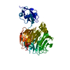
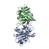
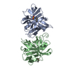
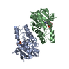
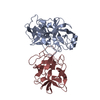
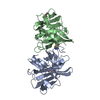
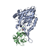
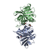
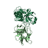
Crystal structure of Rea1-MIDAS/Rsa4-UBL complex from Chaetomium thermophilum
Components
  • Midasin,Midasin
  • Ribosome assembly protein 4
KeywordsRIBOSOME / ribosome biogenesis / integrin / midas
Function / homology
Function and homology information


ribosome biogenesis / nucleolus / ATP hydrolysis activity / nucleoplasm / ATP binding
Similarity search - Function
Midasin / Midasin, AAA lid domain 7 / Midasin AAA lid domain 5 / : / Midasin AAA lid domain / Midasin AAA lid domain / Midasin AAA+ ATPase lid domain / ATPase, dynein-related, AAA domain / AAA domain (dynein-related subfamily) / von Willebrand factor (vWF) type A domain ...Midasin / Midasin, AAA lid domain 7 / Midasin AAA lid domain 5 / : / Midasin AAA lid domain / Midasin AAA lid domain / Midasin AAA+ ATPase lid domain / ATPase, dynein-related, AAA domain / AAA domain (dynein-related subfamily) / von Willebrand factor (vWF) type A domain / VWFA domain profile. / von Willebrand factor, type A / von Willebrand factor A-like domain superfamily / G-protein beta WD-40 repeat / WD40 repeat, conserved site / Trp-Asp (WD) repeats signature. / WD domain, G-beta repeat / WD40 repeats / WD40 repeat / Trp-Asp (WD) repeats profile. / Trp-Asp (WD) repeats circular profile. / WD40-repeat-containing domain superfamily / WD40/YVTN repeat-like-containing domain superfamily / ATPases associated with a variety of cellular activities / AAA+ ATPase domain / P-loop containing nucleoside triphosphate hydrolase
Similarity search - Domain/homology
Ribosome assembly protein 4 / Midasin
Similarity search - Component
Biological speciesChaetomium thermophilum var. thermophilum DSM 1495 (fungus)
MethodX-RAY DIFFRACTION / SYNCHROTRON / MOLECULAR REPLACEMENT / SAD / molecular replacement / Resolution: 1.89 Å
AuthorsAhmed, Y.L. / Thoms, M. / Hurt, E. / Sinning, I.
Funding support Germany, 1items
OrganizationGrant numberCountry
Germany
CitationJournal: Nat Commun / Year: 2019
Title: Crystal structures of Rea1-MIDAS bound to its ribosome assembly factor ligands resembling integrin-ligand-type complexes.
Authors: Ahmed, Y.L. / Thoms, M. / Mitterer, V. / Sinning, I. / Hurt, E.
History
DepositionFeb 22, 2019Deposition site: PDBE / Processing site: PDBE
Revision 1.0Aug 7, 2019Provider: repository / Type: Initial release
Revision 1.1Jan 24, 2024Group: Data collection / Database references ...Data collection / Database references / Derived calculations / Refinement description
Category: chem_comp_atom / chem_comp_bond ...chem_comp_atom / chem_comp_bond / database_2 / pdbx_initial_refinement_model / pdbx_struct_conn_angle / struct_conn
Item: _database_2.pdbx_DOI / _database_2.pdbx_database_accession ..._database_2.pdbx_DOI / _database_2.pdbx_database_accession / _pdbx_struct_conn_angle.ptnr1_auth_asym_id / _pdbx_struct_conn_angle.ptnr1_auth_comp_id / _pdbx_struct_conn_angle.ptnr1_auth_seq_id / _pdbx_struct_conn_angle.ptnr1_label_asym_id / _pdbx_struct_conn_angle.ptnr1_label_atom_id / _pdbx_struct_conn_angle.ptnr1_label_comp_id / _pdbx_struct_conn_angle.ptnr1_label_seq_id / _pdbx_struct_conn_angle.ptnr3_auth_asym_id / _pdbx_struct_conn_angle.ptnr3_auth_comp_id / _pdbx_struct_conn_angle.ptnr3_auth_seq_id / _pdbx_struct_conn_angle.ptnr3_label_asym_id / _pdbx_struct_conn_angle.ptnr3_label_atom_id / _pdbx_struct_conn_angle.ptnr3_label_comp_id / _pdbx_struct_conn_angle.ptnr3_label_seq_id / _pdbx_struct_conn_angle.value / _struct_conn.pdbx_dist_value / _struct_conn.ptnr1_auth_asym_id / _struct_conn.ptnr1_auth_comp_id / _struct_conn.ptnr1_auth_seq_id / _struct_conn.ptnr1_label_asym_id / _struct_conn.ptnr1_label_atom_id / _struct_conn.ptnr1_label_comp_id / _struct_conn.ptnr1_label_seq_id / _struct_conn.ptnr2_auth_asym_id / _struct_conn.ptnr2_auth_comp_id / _struct_conn.ptnr2_auth_seq_id / _struct_conn.ptnr2_label_asym_id / _struct_conn.ptnr2_label_atom_id / _struct_conn.ptnr2_label_comp_id / _struct_conn.ptnr2_label_seq_id

-
Structure visualization

Structure viewerMolecule:
MolmilJmol/JSmol

Downloads & links

-
Assembly

Deposited unit
A: Midasin,Midasin
B: Ribosome assembly protein 4
hetero molecules


Theoretical massNumber of molelcules
Total (without water)44,74613
Polymers43,7892
Non-polymers95711
Water3,945219
1


  • Idetical with deposited unit
  • defined by author&software
  • Evidence: gel filtration
TypeNameSymmetry operationNumber
identity operation1_555x,y,z1
Buried area4100 Å2
ΔGint-52 kcal/mol
Surface area17250 Å2
MethodPISA
Unit cell
Length a, b, c (Å)115.675, 115.675, 74.623
Angle α, β, γ (deg.)90.000, 90.000, 90.000
Int Tables number96
Space group name H-MP43212
Components on special symmetry positions
IDModelComponents
11A-5207-

HOH

21A-5332-

HOH

-
Components

-
Protein , 2 types, 2 molecules AB

#1: Protein Midasin,Midasin


Mass: 31464.826 Da / Num. of mol.: 1
Source method: isolated from a genetically manipulated source
Source: (gene. exp.) Chaetomium thermophilum var. thermophilum DSM 1495 (fungus)
Gene: CTHT_0069750 / Production host: Escherichia coli BL21(DE3) (bacteria) / References: UniProt: G0SHE6
#2: Protein Ribosome assembly protein 4 / Notchless protein homolog 1 / Ribosome biogenesis factor RSA4


Mass: 12323.778 Da / Num. of mol.: 1
Source method: isolated from a genetically manipulated source
Source: (gene. exp.) Chaetomium thermophilum var. thermophilum DSM 1495 (fungus)
Gene: CTHT_0055700 / Production host: Escherichia coli BL21(DE3) (bacteria) / References: UniProt: G0SC29

-
Non-polymers , 4 types, 230 molecules

#3: Chemical ChemComp-MG / MAGNESIUM ION


Mass: 24.305 Da / Num. of mol.: 1 / Source method: obtained synthetically / Formula: Mg
#4: Chemical ChemComp-SO4 / SULFATE ION / Sulfate


Mass: 96.063 Da / Num. of mol.: 3 / Source method: obtained synthetically / Formula: SO4
#5: Chemical
ChemComp-GOL / GLYCEROL / GLYCERIN / PROPANE-1,2,3-TRIOL / Glycerol


Mass: 92.094 Da / Num. of mol.: 7 / Source method: obtained synthetically / Formula: C3H8O3
#6: Water ChemComp-HOH / water / Water


Mass: 18.015 Da / Num. of mol.: 219 / Source method: isolated from a natural source / Formula: H2O

-
Experimental details

-
Experiment

ExperimentMethod: X-RAY DIFFRACTION / Number of used crystals: 1

-
Sample preparation

CrystalDensity Matthews: 2.85 Å3/Da / Density % sol: 56.85 %
Crystal growTemperature: 291 K / Method: vapor diffusion, sitting drop / pH: 6.5 / Details: 2 M (NH4)2SO4, 5% PEG 400 and 0.1 M MES at pH 6.5

-
Data collection

DiffractionMean temperature: 100 K / Serial crystal experiment: N
Diffraction sourceSource: SYNCHROTRON / Site: ESRF / Beamline: ID23-2 / Wavelength: 0.8729 Å
DetectorType: DECTRIS PILATUS3 2M / Detector: PIXEL / Date: Feb 5, 2016
RadiationProtocol: SINGLE WAVELENGTH / Monochromatic (M) / Laue (L): M / Scattering type: x-ray
Radiation wavelengthWavelength: 0.8729 Å / Relative weight: 1
ReflectionResolution: 1.89→36.58 Å / Num. obs: 41048 / % possible obs: 99.97 % / Redundancy: 13.2 % / Biso Wilson estimate: 29.28 Å2 / CC1/2: 0.999 / Net I/σ(I): 12.86
Reflection shellResolution: 1.89→1.95 Å / CC1/2: 0.478

-
Phasing

Phasing
Method
SAD
molecular replacement

-
Processing

Software
NameVersionClassification
XDSdata reduction
Aimlessdata scaling
MOLREPphasing
SHELXDEphasing
PHENIX1.12_2829refinement
PDB_EXTRACT3.24data extraction
RefinementMethod to determine structure: MOLECULAR REPLACEMENT
Starting model: 6QT8 and 4WJS (UBL domain only)
Resolution: 1.89→36.58 Å / SU ML: 0.23 / Cross valid method: THROUGHOUT / σ(F): 0.25 / Phase error: 21.76
RfactorNum. reflection% reflection
Rfree0.2114 4020 5.19 %
Rwork0.1758 --
obs0.1776 77444 99.99 %
Solvent computationShrinkage radii: 0.9 Å / VDW probe radii: 1.11 Å
Displacement parametersBiso max: 124.53 Å2 / Biso mean: 42.7495 Å2 / Biso min: 16.94 Å2
Refinement stepCycle: final / Resolution: 1.89→36.58 Å
ProteinNucleic acidLigandSolventTotal
Num. atoms2837 0 58 219 3114
Biso mean--68.99 50.51 -
Num. residues----359
Refine LS restraints
Refine-IDTypeDev idealNumber
X-RAY DIFFRACTIONf_bond_d0.0182965
X-RAY DIFFRACTIONf_angle_d1.4124011
X-RAY DIFFRACTIONf_chiral_restr0.09455
X-RAY DIFFRACTIONf_plane_restr0.008517
X-RAY DIFFRACTIONf_dihedral_angle_d12.6662259
LS refinement shell

Refine-ID: X-RAY DIFFRACTION / Rfactor Rfree error: 0 / Total num. of bins used: 29 / % reflection obs: 100 %

Resolution (Å)Rfactor RfreeNum. reflection RfreeRfactor RworkNum. reflection RworkNum. reflection all
1.89-1.91230.37491080.330125762684
1.9123-1.93560.33991380.324925132651
1.9356-1.96010.34281480.307625432691
1.9601-1.98590.28081570.283724712628
1.9859-2.01310.35121300.266525762706
2.0131-2.04190.28131570.259824932650
2.0419-2.07230.30321480.240125462694
2.0723-2.10470.2391290.222925082637
2.1047-2.13920.24641300.226825532683
2.1392-2.17610.27241560.21825202676
2.1761-2.21570.2271470.197525312678
2.2157-2.25830.20891270.190225452672
2.2583-2.30440.22351150.179625662681
2.3044-2.35450.21881490.182124972646
2.3545-2.40930.23031370.172925832720
2.4093-2.46950.21151390.172424932632
2.4695-2.53630.24811040.173125462650
2.5363-2.61090.23471280.1825482676
2.6109-2.69520.21131440.16825402684
2.6952-2.79150.23651500.168725122662
2.7915-2.90320.19741470.169125292676
2.9032-3.03530.20351360.157425412677
3.0353-3.19530.15681460.158425252671
3.1953-3.39540.19871790.153124872666
3.3954-3.65750.19151420.14825232665
3.6575-4.02540.17831180.137325592677
4.0254-4.60740.14121100.13425792689
4.6074-5.8030.22821510.16225092660
5.803-45.72760.19521500.195425122662
Refinement TLS params.Method: refined / Origin x: 11.9677 Å / Origin y: 36.4946 Å / Origin z: 5.2227 Å
111213212223313233
T0.0725 Å20.0032 Å2-0.0015 Å2-0.1235 Å20.0118 Å2--0.1309 Å2
L1.3445 °2-0.5687 °2-0.2272 °2-1.2738 °2-0.0029 °2--1.6024 °2
S-0.0522 Å °-0.1587 Å °0.0428 Å °0.0072 Å °0.0681 Å °0.1091 Å °-0.057 Å °-0.0046 Å °-0.0004 Å °
Refinement TLS group
IDRefine-IDRefine TLS-IDSelection detailsAuth asym-IDAuth seq-ID
1X-RAY DIFFRACTION1allA4696 - 4997
2X-RAY DIFFRACTION1allB28 - 128
3X-RAY DIFFRACTION1allC1
4X-RAY DIFFRACTION1allD1 - 3
5X-RAY DIFFRACTION1allG5001 - 5601
6X-RAY DIFFRACTION1allS1 - 219

+
About Yorodumi

-
News

-
Feb 9, 2022. New format data for meta-information of EMDB entries

New format data for meta-information of EMDB entries

  • Version 3 of the EMDB header file is now the official format.
  • The previous official version 1.9 will be removed from the archive.

Related info.:EMDB header

External links:wwPDB to switch to version 3 of the EMDB data model

-
Aug 12, 2020. Covid-19 info

Covid-19 info

URL: https://pdbj.org/emnavi/covid19.php

New page: Covid-19 featured information page in EM Navigator.

Related info.:Covid-19 info / Mar 5, 2020. Novel coronavirus structure data

+
Mar 5, 2020. Novel coronavirus structure data

Novel coronavirus structure data

Related info.:Yorodumi Speices / Aug 12, 2020. Covid-19 info

External links:COVID-19 featured content - PDBj / Molecule of the Month (242):Coronavirus Proteases

+
Jan 31, 2019. EMDB accession codes are about to change! (news from PDBe EMDB page)

EMDB accession codes are about to change! (news from PDBe EMDB page)

  • The allocation of 4 digits for EMDB accession codes will soon come to an end. Whilst these codes will remain in use, new EMDB accession codes will include an additional digit and will expand incrementally as the available range of codes is exhausted. The current 4-digit format prefixed with “EMD-” (i.e. EMD-XXXX) will advance to a 5-digit format (i.e. EMD-XXXXX), and so on. It is currently estimated that the 4-digit codes will be depleted around Spring 2019, at which point the 5-digit format will come into force.
  • The EM Navigator/Yorodumi systems omit the EMD- prefix.

Related info.:Q: What is EMD? / ID/Accession-code notation in Yorodumi/EM Navigator

External links:EMDB Accession Codes are Changing Soon! / Contact to PDBj

+
Jul 12, 2017. Major update of PDB

Major update of PDB

  • wwPDB released updated PDB data conforming to the new PDBx/mmCIF dictionary.
  • This is a major update changing the version number from 4 to 5, and with Remediation, in which all the entries are updated.
  • In this update, many items about electron microscopy experimental information are reorganized (e.g. em_software).
  • Now, EM Navigator and Yorodumi are based on the updated data.

External links:wwPDB Remediation / Enriched Model Files Conforming to OneDep Data Standards Now Available in the PDB FTP Archive

-
Yorodumi

Thousand views of thousand structures

  • Yorodumi is a browser for structure data from EMDB, PDB, SASBDB, etc.
  • This page is also the successor to EM Navigator detail page, and also detail information page/front-end page for Omokage search.
  • The word "yorodu" (or yorozu) is an old Japanese word meaning "ten thousand". "mi" (miru) is to see.

Related info.:EMDB / PDB / SASBDB / Comparison of 3 databanks / Yorodumi Search / Aug 31, 2016. New EM Navigator & Yorodumi / Yorodumi Papers / Jmol/JSmol / Function and homology information / Changes in new EM Navigator and Yorodumi

Read more