ムービー
ムービー コントローラー


+ データを開く

- 基本情報
基本情報
| 登録情報 | データベース: PDB / ID: 1ld4 | ||||||
|---|---|---|---|---|---|---|---|
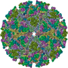
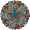
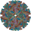
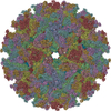
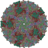
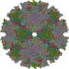
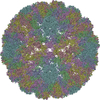
| タイトル | Placement of the Structural Proteins in Sindbis Virus | ||||||
 要素 要素 |
| ||||||
 キーワード キーワード | VIRUS / sindbis virus / alphavirus structure / glycoprotein organization / nucleocapsid structure / transmembrane coiled coils / Icosahedral virus | ||||||
| 機能・相同性 |  機能・相同性情報 機能・相同性情報icosahedral viral capsid, spike / FCERI mediated MAPK activation / protein localization to nuclear periphery / Activation of the AP-1 family of transcription factors / response to amino acid starvation / negative regulation of ribosomal protein gene transcription by RNA polymerase II / positive regulation of cellular response to amino acid starvation / mediator complex binding / togavirin / Oxidative Stress Induced Senescence ...icosahedral viral capsid, spike / FCERI mediated MAPK activation / protein localization to nuclear periphery / Activation of the AP-1 family of transcription factors / response to amino acid starvation / negative regulation of ribosomal protein gene transcription by RNA polymerase II / positive regulation of cellular response to amino acid starvation / mediator complex binding / togavirin / Oxidative Stress Induced Senescence / T=4 icosahedral viral capsid / TFIID-class transcription factor complex binding / ubiquitin-like protein ligase binding / amino acid biosynthetic process / positive regulation of RNA polymerase II transcription preinitiation complex assembly / positive regulation of transcription initiation by RNA polymerase II / cellular response to nutrient levels / cellular response to amino acid starvation / RNA polymerase II transcription regulator complex / DNA-binding transcription activator activity, RNA polymerase II-specific / symbiont-mediated suppression of host toll-like receptor signaling pathway / transcription regulator complex / clathrin-dependent endocytosis of virus by host cell / sequence-specific DNA binding / RNA polymerase II-specific DNA-binding transcription factor binding / membrane fusion / DNA-binding transcription factor activity, RNA polymerase II-specific / host cell cytoplasm / intracellular signal transduction / RNA polymerase II cis-regulatory region sequence-specific DNA binding / DNA-binding transcription factor activity / viral translational frameshifting / serine-type endopeptidase activity / fusion of virus membrane with host endosome membrane / viral envelope / chromatin binding / virion attachment to host cell / host cell nucleus / host cell plasma membrane / virion membrane / structural molecule activity / negative regulation of transcription by RNA polymerase II / positive regulation of transcription by RNA polymerase II / proteolysis / RNA binding / identical protein binding / nucleus / membrane 類似検索 - 分子機能 | ||||||
| 生物種 |  Sindbis virus (シンドビスウイルス) Sindbis virus (シンドビスウイルス) | ||||||
| 手法 | 電子顕微鏡法 / 単粒子再構成法 / クライオ電子顕微鏡法 / 解像度: 11.4 Å | ||||||
 データ登録者 データ登録者 | Zhang, W. / Mukhopadhyay, S. / Pletnev, S.V. / Baker, T.S. / Kuhn, R.J. / Rossmann, M.G. | ||||||
 引用 引用 | ジャーナル: J Virol / 年: 2002 タイトル: Placement of the structural proteins in Sindbis virus. 著者: Wei Zhang / Suchetana Mukhopadhyay / Sergei V Pletnev / Timothy S Baker / Richard J Kuhn / Michael G Rossmann /  要旨: The structure of the lipid-enveloped Sindbis virus has been determined by fitting atomic resolution crystallographic structures of component proteins into an 11-A resolution cryoelectron microscopy ...The structure of the lipid-enveloped Sindbis virus has been determined by fitting atomic resolution crystallographic structures of component proteins into an 11-A resolution cryoelectron microscopy map. The virus has T=4 quasisymmetry elements that are accurately maintained between the external glycoproteins, the transmembrane helical region, and the internal nucleocapsid core. The crystal structure of the E1 glycoprotein was fitted into the cryoelectron microscopy density, in part by using the known carbohydrate positions as restraints. A difference map showed that the E2 glycoprotein was shaped similarly to E1, suggesting a possible common evolutionary origin for these two glycoproteins. The structure shows that the E2 glycoprotein would have to move away from the center of the trimeric spike in order to expose enough viral membrane surface to permit fusion with the cellular membrane during the initial stages of host infection. The well-resolved E1-E2 transmembrane regions form alpha-helical coiled coils that were consistent with T=4 symmetry. The known structure of the capsid protein was fitted into the density corresponding to the nucleocapsid, revising the structure published earlier. | ||||||
| 履歴 |
| ||||||
| Remark 99 | Chains E-L contain alpha-carbons only. The C-terminal residues of chains M-P contain alpha-carbons only. | ||||||
| Remark 600 | HETEROGEN THE COORDINATES OF UNX 1139, 1141, 1245, 1196, 1200, 1262, 1318, 4139, 4141, 4245, 4196, ...HETEROGEN THE COORDINATES OF UNX 1139, 1141, 1245, 1196, 1200, 1262, 1318, 4139, 4141, 4245, 4196, 4200, 4262, 4318, 5139, 5141, 5245, 5196, 5200, 5262, 5318, 6139, 6141, 6245, 6196, 6200, 6262, 6318 ARE THE CENTERS OF DIFFERENCE DENSITY PEAKS THAT CORRESPOND TO THE CARBOHYDRATE MOLECULES OF THE VIRION. THE COORDINATES OF UNX 1000, 4000, 5000, AND 6000 ARE THE POINTS WHERE THE GLYCOPROTEINS E1 AND E2 ENTERS INTO MEMBRANE. |
- 構造の表示
構造の表示
| ムービー |
ムービービューア |
|---|---|
| 構造ビューア | 分子: MolmilJmol/JSmol |
- ダウンロードとリンク
-ダウンロード
| PDBx/mmCIF形式 | 1ld4.cif.gz | 420.9 KB | 表示 | PDBx/mmCIF形式 |
|---|---|---|---|---|
| PDB形式 | pdb1ld4.ent.gz | 322.5 KB | 表示 | PDB形式 |
| PDBx/mmJSON形式 | 1ld4.json.gz | ツリー表示 | PDBx/mmJSON形式 | |
| その他 |  その他のダウンロード その他のダウンロード |
-検証レポート
| 文書・要旨 | 1ld4_validation.pdf.gz | 417.2 KB | 表示 | wwPDB検証レポート |
|---|---|---|---|---|
| 文書・詳細版 | 1ld4_full_validation.pdf.gz | 458.9 KB | 表示 | |
| XML形式データ | 1ld4_validation.xml.gz | 40.2 KB | 表示 | |
| CIF形式データ | 1ld4_validation.cif.gz | 59.5 KB | 表示 | |
| アーカイブディレクトリ | https://data.pdbj.org/pub/pdb/validation_reports/ld/1ld4ftp://data.pdbj.org/pub/pdb/validation_reports/ld/1ld4 | HTTPS FTP |
-関連構造データ
| 関連構造データ | |
|---|---|
| 類似構造データ |


-リンク
 PDBj
PDBj


- 集合体
集合体
| 登録構造単位 | 
|
|---|---|
| 1 | x 60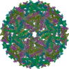
|
| 2 |
|
| 3 | x 5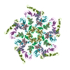
|
| 4 | x 6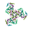
|
| 5 | 
|
| 対称性 | 点対称性: (ヘルマン・モーガン記号: 532 / シェーンフリース記号: I (正20面体型対称)) |
-要素
| #1: タンパク質 | 分子量: 29410.010 Da / 分子数: 4 / 由来タイプ: 天然 由来: (天然) Sindbis virus (シンドビスウイルス)属: Alphavirus 参照: UniProt: P03316, 加水分解酵素; プロテアーゼ; ペプチド結合加水分解酵素; セリンエンドペプチターゼ #2: タンパク質 | 分子量: 6718.774 Da / 分子数: 8 / Fragment: LEUCINE-ZIPPER (residues 225-281) / 由来タイプ: 天然 / 由来: (天然) #3: タンパク質 | 分子量: 47416.836 Da / 分子数: 4 / 由来タイプ: 天然 由来: (天然) Sindbis virus (シンドビスウイルス)属: Alphavirus / 参照: UniProt: P03316 #4: 化合物 | ChemComp-UNX /   分子数: 32 / 由来タイプ: 合成 分子数: 32 / 由来タイプ: 合成Has protein modification | Y | |
|---|
-実験情報
-実験
| 実験 | 手法: 電子顕微鏡法 |
|---|---|
| EM実験 | 試料の集合状態: PARTICLE / 3次元再構成法: 単粒子再構成法 |
- 試料調製
試料調製
| 構成要素 | 名称: Sindbis Virus / タイプ: VIRUS |
|---|---|
| ウイルスについての詳細 | ホストのカテゴリ: VERTEBRATES / タイプ: VIRION |
| 天然宿主 | 生物種: Homo sapiens |
| 緩衝液 | 名称: 50mM Tris-HCl, 200mM NaCl, 0.1mM EDTA / pH: 7.5 / 詳細: 50mM Tris-HCl, 200mM NaCl, 0.1mM EDTA |
| 試料 | 濃度: 6 mg/ml / 包埋: NO / シャドウイング: NO / 染色: NO / 凍結: YES |
| 結晶化 | *PLUS 手法: cryo-electron microscopy / 詳細: cryo-electron microscopy |
- 電子顕微鏡撮影
| 顕微鏡 | モデル: FEI/PHILIPS CM200T / 日付: 2000年6月15日 |
|---|---|
| 電子銃 | 電子線源:  FIELD EMISSION GUN / 加速電圧: 200 kV / 照射モード: FLOOD BEAM FIELD EMISSION GUN / 加速電圧: 200 kV / 照射モード: FLOOD BEAM |
| 電子レンズ | モード: BRIGHT FIELD / 倍率(公称値): 38000 X / 最大 デフォーカス(公称値): 2580 nm / 最小 デフォーカス(公称値): 1100 nm / Cs: 2 mm |
| 試料ホルダ | 温度: 87 K / 傾斜角・最大: 0 ° / 傾斜角・最小: 0 ° |
| 撮影 | 電子線照射量: 18.4 e/Å2 / フィルム・検出器のモデル: KODAK SO-163 FILM 詳細: films were developed in full-strength of Kodak D19 developer for 12 minutes at 20 C |
- 解析
解析
| EMソフトウェア |
| ||||||||||||||||||||||||||||
|---|---|---|---|---|---|---|---|---|---|---|---|---|---|---|---|---|---|---|---|---|---|---|---|---|---|---|---|---|---|
| CTF補正 | 詳細: each viral image was CTF corrected before reconstruction, based on the following equation: F(corr)=F(obs)/[|CTF|+wiener] | ||||||||||||||||||||||||||||
| 対称性 | 点対称性: I (正20面体型対称) | ||||||||||||||||||||||||||||
| 3次元再構成 | 手法: FOURIER-BESSEL method Reference: Baker, T. S., N. H. Olson, and S. D. Fuller (1999). Adding the third dimension to virus life cycles: Three-dimensional reconstruction of icosahedral viruses ...手法: FOURIER-BESSEL method Reference: Baker, T. S., N. H. Olson, and S. D. Fuller (1999). Adding the third dimension to virus life cycles: Three-dimensional reconstruction of icosahedral viruses from cryo-electron micrographs. Micro. & Mol. Bio. Rev. 63:862-922. 解像度: 11.4 Å / ピクセルサイズ(公称値): 3.68 Å / 対称性のタイプ: POINT | ||||||||||||||||||||||||||||
| 原子モデル構築 | B value: 1000 / 空間: REAL 詳細: DETAILS--The structure of the lipid-enveloped Sindbis virus has been determined by fitting atomic resolution crystallographic structures of component proteins into an 11 resolution cryo- ...詳細: DETAILS--The structure of the lipid-enveloped Sindbis virus has been determined by fitting atomic resolution crystallographic structures of component proteins into an 11 resolution cryo- electron microscopy (cryoEM) map (with significant terms to 10 resolution). The E1 glycoprotein, whose structure had been determined independently for the homologous Semliki Forest virus by X-ray crystallography (Lescar, J., Roussel,A., Wein, M.W., Navaza, J., Fuller, S.D., Wengler, G., and Rey, F.A. (2001). The fusion glycoprotein shell of Semliki Forest virus- an icosahedral assembly primed for fusogenic activation at endosomal pH. Cell 105, 137-148), was fitted into the cryoEM density, in part by using the known positions of glycoprotein sites as restraints. The well-resolved transmembrane regions form coiled coils that could be fitted with the dimeric GCN4 coiled-coil structure. The helices could be seen to extend to E1 and E2 on the outside of the membrane and to the nucleocapsid on the inside. The known structure of the capsid protein (Choi, H.K., Tong, L., Minor, W., Dumas, P., Boege, U., Rossmann, M.G., and Wengler, G. (1991). Structure of Sindbis virus core protein reveals a chymotrypsin-like serine proteinase and the organization of the virion. Nature 354, 37-43.) was fitted into the density corresponding to the nucleocapsid, revising an earlier published structure. | ||||||||||||||||||||||||||||
| 原子モデル構築 |
| ||||||||||||||||||||||||||||
| 精密化 | 最高解像度: 11.4 Å | ||||||||||||||||||||||||||||
| 精密化ステップ | サイクル: LAST / 最高解像度: 11.4 Å
|