 Movie
Movie Controller


+ Open data

- Basic information
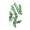
| Entry | Database: PDB / ID: 1krq | ||||||
|---|---|---|---|---|---|---|---|
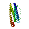
| Title | CRYSTAL STRUCTURE ANALYSIS OF CAMPYLOBACTER JEJUNI FERRITIN | ||||||
 Components Components | ferritin | ||||||
 Keywords Keywords | METAL BINDING PROTEIN / bacterial non-heme ferritin / H-chain like four-helix bundle | ||||||
| Function / homology |  Function and homology information Function and homology informationbacterial non-heme ferritin / ferroxidase activity / ferric iron binding / iron ion transport / ferrous iron binding / intracellular iron ion homeostasis / cytosol Similarity search - Function | ||||||
| Biological species |   Campylobacter jejuni (Campylobacter) Campylobacter jejuni (Campylobacter) | ||||||
| Method |  X-RAY DIFFRACTION / SYNCHROTRON / MOLECULAR REPLACEMENT / Resolution: 2.7 Å X-RAY DIFFRACTION / SYNCHROTRON / MOLECULAR REPLACEMENT / Resolution: 2.7 Å | ||||||
 Authors Authors | Hortolan, L. / Saintout, N. / Granier, G. / Langlois d'Estaintot, B. / Manigand, C. / Mizunoe, Y. / Wai, S.N. / Gallois, B. / Precigoux, G. | ||||||
 Citation Citation | Journal: To be Published Title: STRUCTURE OF CAMPYLOBACTER JEJUNI FERRITIN AT 2.7 A RESOLUTION Authors: Hortolan, L. / Saintout, N. / Granier, G. / Langlois d'Estaintot, B. / Manigand, C. / Mizunoe, Y. / Wai, S.N. / Gallois, B. / Precigoux, G. | ||||||
| History |
| ||||||
| Remark 525 | SOLVENT WATER MOLECULES HOH 200 AND HOH 201 BIND THE PROTEIN AT THE FERROXYDASE CENTER (INVOLVED ...SOLVENT WATER MOLECULES HOH 200 AND HOH 201 BIND THE PROTEIN AT THE FERROXYDASE CENTER (INVOLVED RESIDUES: GLU 17, GLU 50, HIS 53, GLU 94, GLU 126, GLU 129 and GLU 130) PREVIOUSLY DESCRIBED IN THE FERRITIN OF E.Coli (PDB entry code 1EUM) AND THUS COULD STAND FOR IRON IONS WITH LOW OCCUPANCY FACTORS. |
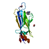
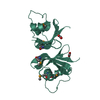
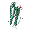
- Structure visualization
| Structure viewer | Molecule: MolmilJmol/JSmol |
|---|
- Downloads & links
-Download
| PDBx/mmCIF format | 1krq.cif.gz | 45.4 KB | Display | PDBx/mmCIF format |
|---|---|---|---|---|
| PDB format | pdb1krq.ent.gz | 32.5 KB | Display | PDB format |
| PDBx/mmJSON format | 1krq.json.gz | Tree view | PDBx/mmJSON format | |
| Others |  Other downloads Other downloads |
-Validation report
| Summary document | 1krq_validation.pdf.gz | 424.8 KB | Display | wwPDB validaton report |
|---|---|---|---|---|
| Full document | 1krq_full_validation.pdf.gz | 425.9 KB | Display | |
| Data in XML | 1krq_validation.xml.gz | 7.7 KB | Display | |
| Data in CIF | 1krq_validation.cif.gz | 9.4 KB | Display | |
| Arichive directory | https://data.pdbj.org/pub/pdb/validation_reports/kr/1krqftp://data.pdbj.org/pub/pdb/validation_reports/kr/1krq | HTTPS FTP |
-Related structure data
| Related structure data |  2fhaS S: Starting model for refinement |
|---|---|
| Similar structure data |

-Links
 PDBj
PDBj


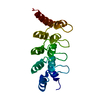
- Assembly
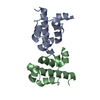
| Deposited unit | 
| ||||||||
|---|---|---|---|---|---|---|---|---|---|
| 1 |
| ||||||||
| 2 | x 24
| ||||||||
| Unit cell |
|
-Components
| #1: Protein | Mass: 19510.986 Da / Num. of mol.: 1 Source method: isolated from a genetically manipulated source Source: (gene. exp.) Campylobacter jejuni (Campylobacter) / Gene: cft / Plasmid: pET3b / Species (production host): Escherichia coli / Production host: |
|---|---|
| #2: Water | ChemComp-HOH /  Mass: 18.015 Da / Num. of mol.: 4 / Source method: isolated from a natural source / Formula: H2O Mass: 18.015 Da / Num. of mol.: 4 / Source method: isolated from a natural source / Formula: H2O |
-Experimental details
-Experiment
| Experiment | Method: X-RAY DIFFRACTION / Number of used crystals: 1 |
|---|
- Sample preparation
| Crystal | Density Matthews: 3.95 Å3/Da / Density % sol: 68.62 % |
|---|---|
| Crystal grow | Temperature: 300 K / Method: vapor diffusion, hanging drop / pH: 7.5 Details: Ammonium sulfate, PEG400, Hepes, Sodium azide, pH 7.5, VAPOR DIFFUSION, HANGING DROP, temperature 300K |
-Data collection
| Diffraction | Mean temperature: 296 K |
|---|---|
| Diffraction source | Source: SYNCHROTRON / Site: LURE  / Beamline: DW32 / Wavelength: 0.966 Å / Beamline: DW32 / Wavelength: 0.966 Å |
| Detector | Type: MARRESEARCH / Detector: IMAGE PLATE / Date: Apr 17, 2000 / Details: W/Si mutilayer mirror |
| Radiation | Monochromator: focused Si(111) / Protocol: SINGLE WAVELENGTH / Monochromatic (M) / Laue (L): M / Scattering type: x-ray |
| Radiation wavelength | Wavelength: 0.966 Å / Relative weight: 1 |
| Reflection | Resolution: 2.7→19 Å / Num. all: 8373 / Num. obs: 8373 / % possible obs: 98.5 % / Observed criterion σ(I): 0 / Redundancy: 6.5 % / Biso Wilson estimate: 44.75 Å2 / Rsym value: 0.064 / Net I/σ(I): 5.9 |
| Reflection shell | Resolution: 2.7→2.77 Å / Redundancy: 5.7 % / Mean I/σ(I) obs: 2 / Rsym value: 0.38 / % possible all: 99.8 |
- Processing
| Software |
| ||||||||||||||||||||||||||||||||||||||||||||||||||||||||||||||||||||||||||||||||
|---|---|---|---|---|---|---|---|---|---|---|---|---|---|---|---|---|---|---|---|---|---|---|---|---|---|---|---|---|---|---|---|---|---|---|---|---|---|---|---|---|---|---|---|---|---|---|---|---|---|---|---|---|---|---|---|---|---|---|---|---|---|---|---|---|---|---|---|---|---|---|---|---|---|---|---|---|---|---|---|---|---|
| Refinement | Method to determine structure: MOLECULAR REPLACEMENT Starting model: PDB ENTRY 2FHA Resolution: 2.7→15 Å / Isotropic thermal model: Isotropic / Cross valid method: THROUGHOUT / σ(F): 0 / Stereochemistry target values: Engh & Huber
| ||||||||||||||||||||||||||||||||||||||||||||||||||||||||||||||||||||||||||||||||
| Solvent computation | Solvent model: CALCULATED FROM COORDINATES / Bsol: 64.48 Å2 / ksol: 0.37 e/Å3 | ||||||||||||||||||||||||||||||||||||||||||||||||||||||||||||||||||||||||||||||||
| Displacement parameters | Biso mean: 53.2 Å2 | ||||||||||||||||||||||||||||||||||||||||||||||||||||||||||||||||||||||||||||||||
| Refine analyze |
| ||||||||||||||||||||||||||||||||||||||||||||||||||||||||||||||||||||||||||||||||
| Refinement step | Cycle: LAST / Resolution: 2.7→15 Å
| ||||||||||||||||||||||||||||||||||||||||||||||||||||||||||||||||||||||||||||||||
| Refine LS restraints |
| ||||||||||||||||||||||||||||||||||||||||||||||||||||||||||||||||||||||||||||||||
| LS refinement shell |
|
