 Movie
Movie Controller

[English] 日本語
 Yorodumi
- PDB-2za8: recombinant horse L-chain apoferritin N-terminal deletion mutant ... -
+ Open data

- Basic information
| Entry | Database: PDB / ID: 2za8 | ||||||
|---|---|---|---|---|---|---|---|
| Title | recombinant horse L-chain apoferritin N-terminal deletion mutant (residues 1-8) | ||||||
 Components Components | Ferritin light chain | ||||||
 Keywords Keywords | METAL BINDING PROTEIN / ferric iron binding / Acetylation / Iron storage / Metal-binding | ||||||
| Function / homology |  Function and homology information Function and homology informationferritin complex / autolysosome / ferric iron binding / autophagosome / iron ion transport / ferrous iron binding / cytoplasmic vesicle / intracellular iron ion homeostasis / iron ion binding / cytoplasm Similarity search - Function | ||||||
| Biological species |  | ||||||
| Method |  X-RAY DIFFRACTION / SYNCHROTRON / MOLECULAR REPLACEMENT / Resolution: 1.4 Å X-RAY DIFFRACTION / SYNCHROTRON / MOLECULAR REPLACEMENT / Resolution: 1.4 Å | ||||||
 Authors Authors | Yamashita, I. / Mishima, Y. / Park, S.-Y. / Heddle, J.G. / Tame, J.R.H. | ||||||
 Citation Citation | Journal: J.BIOCHEM.(TOKYO) / Year: 2007 Title: Effect of N-terminal Residues on the Structural Stability of Recombinant Horse L-chain Apoferritin in an Acidic Environment Authors: Yoshizawa, K. / Mishima, Y. / Park, S.-Y. / Heddle, J.G. / Tame, J.R.H. / Iwahori, K. / Kobayashi, M. / Yamashita, I. | ||||||
| History |
|
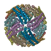
- Structure visualization
| Structure viewer | Molecule: MolmilJmol/JSmol |
|---|
- Downloads & links
-Download
| PDBx/mmCIF format | 2za8.cif.gz | 51.6 KB | Display | PDBx/mmCIF format |
|---|---|---|---|---|
| PDB format | pdb2za8.ent.gz | 37.5 KB | Display | PDB format |
| PDBx/mmJSON format | 2za8.json.gz | Tree view | PDBx/mmJSON format | |
| Others |  Other downloads Other downloads |
-Validation report
| Arichive directory | https://data.pdbj.org/pub/pdb/validation_reports/za/2za8ftp://data.pdbj.org/pub/pdb/validation_reports/za/2za8 | HTTPS FTP |
|---|
-Related structure data
| Related structure data | 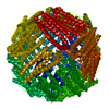 2za6C  2za7C 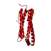 1aewS C: citing same article ( S: Starting model for refinement |
|---|---|
| Similar structure data |

-Links
 PDBj
PDBj


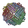
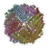
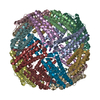
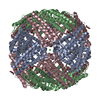
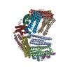
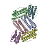
- Assembly
| Deposited unit | 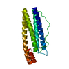
| ||||||||
|---|---|---|---|---|---|---|---|---|---|
| 1 | x 24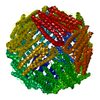
| ||||||||
| Unit cell |
|
-Components
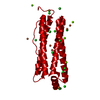
| #1: Protein | Mass: 19041.512 Da / Num. of mol.: 1 / Fragment: residues 9-175 Source method: isolated from a genetically manipulated source Source: (gene. exp.)  | ||||
|---|---|---|---|---|---|
| #2: Chemical |   Mass: 112.411 Da / Num. of mol.: 2 / Source method: obtained synthetically / Formula: Cd Mass: 112.411 Da / Num. of mol.: 2 / Source method: obtained synthetically / Formula: Cd#3: Water | ChemComp-HOH / |  Mass: 18.015 Da / Num. of mol.: 177 / Source method: isolated from a natural source / Formula: H2O Mass: 18.015 Da / Num. of mol.: 177 / Source method: isolated from a natural source / Formula: H2OSequence details | SEE REF. 2 IN THE DATABASE, P02791 IN UNP. | |
-Experimental details
-Experiment
| Experiment | Method: X-RAY DIFFRACTION / Number of used crystals: 1 |
|---|
- Sample preparation
| Crystal | Density Matthews: 3.2 Å3/Da / Density % sol: 61.58 % |
|---|---|
| Crystal grow | Temperature: 293 K / Method: vapor diffusion, hanging drop / pH: 6.8 Details: 0.1M Tris-HCl, 225mM ammonium sulphate, 0.13% cadmium chloride, 25% 2-Metyl-2,4-pentanediol, pH 6.8, VAPOR DIFFUSION, HANGING DROP, temperature 293.0K |
-Data collection
| Diffraction source | Source: SYNCHROTRON / Site: SPring-8  / Beamline: BL44B2 / Wavelength: 0.9 Å / Beamline: BL44B2 / Wavelength: 0.9 Å |
|---|---|
| Detector | Type: MAR CCD 165 mm / Detector: CCD / Date: Oct 20, 2003 |
| Radiation | Protocol: SINGLE WAVELENGTH / Monochromatic (M) / Laue (L): M / Scattering type: x-ray |
| Radiation wavelength | Wavelength: 0.9 Å / Relative weight: 1 |
| Reflection | Resolution: 1.4→20 Å / Num. obs: 52831 / % possible obs: 99.8 % / Biso Wilson estimate: 14 Å2 |
- Processing
| Software |
| ||||||||||||||||||||||||||||||||||||||||||||||||||||||||||||||||||||||||||||||||||||||||||||||||||||||||||||||||||||||||||||||||||||||||||||||||||||||||||||||||||||||||||
|---|---|---|---|---|---|---|---|---|---|---|---|---|---|---|---|---|---|---|---|---|---|---|---|---|---|---|---|---|---|---|---|---|---|---|---|---|---|---|---|---|---|---|---|---|---|---|---|---|---|---|---|---|---|---|---|---|---|---|---|---|---|---|---|---|---|---|---|---|---|---|---|---|---|---|---|---|---|---|---|---|---|---|---|---|---|---|---|---|---|---|---|---|---|---|---|---|---|---|---|---|---|---|---|---|---|---|---|---|---|---|---|---|---|---|---|---|---|---|---|---|---|---|---|---|---|---|---|---|---|---|---|---|---|---|---|---|---|---|---|---|---|---|---|---|---|---|---|---|---|---|---|---|---|---|---|---|---|---|---|---|---|---|---|---|---|---|---|---|---|---|---|
| Refinement | Method to determine structure: MOLECULAR REPLACEMENT Starting model: 1AEW Resolution: 1.4→19.76 Å / Cor.coef. Fo:Fc: 0.935 / Cor.coef. Fo:Fc free: 0.925 / SU B: 1.034 / SU ML: 0.041 / Cross valid method: THROUGHOUT / ESU R: 0.06 / ESU R Free: 0.061 / Stereochemistry target values: MAXIMUM LIKELIHOOD
| ||||||||||||||||||||||||||||||||||||||||||||||||||||||||||||||||||||||||||||||||||||||||||||||||||||||||||||||||||||||||||||||||||||||||||||||||||||||||||||||||||||||||||
| Solvent computation | Ion probe radii: 0.8 Å / Shrinkage radii: 0.8 Å / VDW probe radii: 1.4 Å / Solvent model: BABINET MODEL WITH MASK | ||||||||||||||||||||||||||||||||||||||||||||||||||||||||||||||||||||||||||||||||||||||||||||||||||||||||||||||||||||||||||||||||||||||||||||||||||||||||||||||||||||||||||
| Displacement parameters | Biso mean: 14.328 Å2 | ||||||||||||||||||||||||||||||||||||||||||||||||||||||||||||||||||||||||||||||||||||||||||||||||||||||||||||||||||||||||||||||||||||||||||||||||||||||||||||||||||||||||||
| Refinement step | Cycle: LAST / Resolution: 1.4→19.76 Å
| ||||||||||||||||||||||||||||||||||||||||||||||||||||||||||||||||||||||||||||||||||||||||||||||||||||||||||||||||||||||||||||||||||||||||||||||||||||||||||||||||||||||||||
| Refine LS restraints |
| ||||||||||||||||||||||||||||||||||||||||||||||||||||||||||||||||||||||||||||||||||||||||||||||||||||||||||||||||||||||||||||||||||||||||||||||||||||||||||||||||||||||||||
| LS refinement shell | Resolution: 1.4→1.436 Å / Total num. of bins used: 20 /
|
