[English] 日本語
Yorodumi
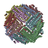
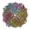
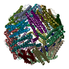
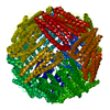
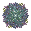
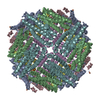
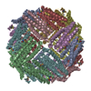
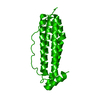
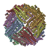
- PDB-3e6r: Crystal structure of apo-ferritin from Pseudo-nitzschia multiseries -

+
Open data


ID or keywords:

Loading...

-
Basic information

Entry
Database: PDB / ID: 3e6r
TitleCrystal structure of apo-ferritin from Pseudo-nitzschia multiseries
ComponentsFerritin
KeywordsOXIDOREDUCTASE / ferritin / apoferritin / iron storage / ferroxidase
Function / homology
Function and homology information


ferroxidase / ferroxidase activity / ferric iron binding / iron ion transport / intracellular iron ion homeostasis / identical protein binding
Similarity search - Function
Ferritin, prokaryotic-type / Ferritin, core subunit, four-helix bundle / Ferritin / Ferritin / Ferritin-like diiron domain / Ferritin-like diiron domain profile. / Ferritin/DPS protein domain / Ferritin-like domain / Ferritin-like / Ferritin-like superfamily ...Ferritin, prokaryotic-type / Ferritin, core subunit, four-helix bundle / Ferritin / Ferritin / Ferritin-like diiron domain / Ferritin-like diiron domain profile. / Ferritin/DPS protein domain / Ferritin-like domain / Ferritin-like / Ferritin-like superfamily / Up-down Bundle / Mainly Alpha
Similarity search - Domain/homology
Biological speciesPseudo-nitzschia multiseries (Diatom)
MethodX-RAY DIFFRACTION / SYNCHROTRON / Resolution: 2.4 Å
AuthorsMurphy, M.E.P. / Arrieta, A.L.
CitationJournal: Nature / Year: 2009
Title: Ferritin is used for iron storage in bloom-forming marine pennate diatoms.
Authors: Marchetti, A. / Parker, M.S. / Moccia, L.P. / Lin, E.O. / Arrieta, A.L. / Ribalet, F. / Murphy, M.E. / Maldonado, M.T. / Armbrust, E.V.
History
DepositionAug 15, 2008Deposition site: RCSB / Processing site: RCSB
Revision 1.0Nov 25, 2008Provider: repository / Type: Initial release
Revision 1.1Jul 13, 2011Group: Version format compliance
Revision 1.2Oct 25, 2017Group: Refinement description / Category: software
Item: _software.classification / _software.contact_author ..._software.classification / _software.contact_author / _software.contact_author_email / _software.date / _software.language / _software.location / _software.name / _software.type / _software.version
Revision 1.3Feb 21, 2024Group: Data collection / Database references / Derived calculations
Category: chem_comp_atom / chem_comp_bond ...chem_comp_atom / chem_comp_bond / database_2 / struct_ref_seq_dif / struct_site
Item: _database_2.pdbx_DOI / _database_2.pdbx_database_accession ..._database_2.pdbx_DOI / _database_2.pdbx_database_accession / _struct_ref_seq_dif.details / _struct_site.pdbx_auth_asym_id / _struct_site.pdbx_auth_comp_id / _struct_site.pdbx_auth_seq_id

-
Structure visualization

Structure viewerMolecule:
MolmilJmol/JSmol

Downloads & links

-
Assembly

Deposited unit
A: Ferritin
B: Ferritin
C: Ferritin
D: Ferritin
E: Ferritin
F: Ferritin
hetero molecules


Theoretical massNumber of molelcules
Total (without water)113,0528
Polymers112,8606
Non-polymers1922
Water12,899716
1
A: Ferritin
B: Ferritin
C: Ferritin
D: Ferritin
E: Ferritin
F: Ferritin
hetero molecules

A: Ferritin
B: Ferritin
C: Ferritin
D: Ferritin
E: Ferritin
F: Ferritin
hetero molecules

A: Ferritin
B: Ferritin
C: Ferritin
D: Ferritin
E: Ferritin
F: Ferritin
hetero molecules

A: Ferritin
B: Ferritin
C: Ferritin
D: Ferritin
E: Ferritin
F: Ferritin
hetero molecules


Theoretical massNumber of molelcules
Total (without water)452,20732
Polymers451,43824
Non-polymers7698
Water43224
TypeNameSymmetry operationNumber
identity operation1_555x,y,z1
crystal symmetry operation2_655-x+1,-y,z1
crystal symmetry operation3_545-y+1/2,x-1/2,z1
crystal symmetry operation4_555y+1/2,-x+1/2,z1
Buried area67050 Å2
ΔGint-438 kcal/mol
Surface area137820 Å2
MethodPISA
Unit cell
Length a, b, c (Å)126.572, 126.572, 170.700
Angle α, β, γ (deg.)90.000, 90.000, 90.000
Int Tables number90
Space group name H-MP4212
Components on special symmetry positions
IDModelComponents
11C-347-

HOH

21D-262-

HOH

31F-286-

HOH

-
Components

#1: Protein
Ferritin /


Mass: 18809.930 Da / Num. of mol.: 6
Source method: isolated from a genetically manipulated source
Source: (gene. exp.) Pseudo-nitzschia multiseries (Diatom) / Strain: CLN 47 / Gene: FTN / Plasmid: pET28a / Production host: Escherichia coli (E. coli) / Strain (production host): BL 21 (DE3) / References: UniProt: B6DMH6, ferroxidase
#2: Chemical ChemComp-SO4 / SULFATE ION / Sulfate


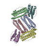
Mass: 96.063 Da / Num. of mol.: 2 / Source method: obtained synthetically / Formula: SO4
#3: Water ChemComp-HOH / water / Water


Mass: 18.015 Da / Num. of mol.: 716 / Source method: isolated from a natural source / Formula: H2O

-
Experimental details

-
Experiment

ExperimentMethod: X-RAY DIFFRACTION / Number of used crystals: 1

-
Sample preparation

CrystalDensity Matthews: 3.03 Å3/Da / Density % sol: 59.39 %
Crystal growTemperature: 293 K / Method: vapor diffusion, hanging drop / pH: 5.5
Details: 1.4 M ammonium sulfate and 0.1 M ammonium acetate pH 5.5, VAPOR DIFFUSION, HANGING DROP, temperature 293K

-
Data collection

DiffractionMean temperature: 90 K
Diffraction sourceSource: SYNCHROTRON / Site: SSRL / Beamline: BL7-1 / Wavelength: 0.9761 Å
DetectorType: ADSC QUANTUM 315 / Detector: CCD / Date: Feb 7, 2008
Details: Vertical focusing mirror; single crystal (Si111) bent monochromator (horizontal focusing).
RadiationMonochromator: side scattering I-beam bent single crystal; asymmetric cut 4.9650 deg.
Protocol: SINGLE WAVELENGTH / Monochromatic (M) / Laue (L): M / Scattering type: x-ray
Radiation wavelengthWavelength: 0.9761 Å / Relative weight: 1
ReflectionResolution: 2.4→100 Å / Num. all: 54922 / Num. obs: 52007 / % possible obs: 94.7 % / Redundancy: 6.4 % / Biso Wilson estimate: 25.9 Å2 / Rmerge(I) obs: 0.095 / Χ2: 1.374
Reflection shell
Resolution (Å)Redundancy (%)Rmerge(I) obsNum. unique allΧ2% possible all
2.4-2.496.40.43652321.3896.8
2.49-2.596.40.35752071.30396.5
2.59-2.76.50.27951961.28696.2
2.7-2.856.50.20452061.27896
2.85-3.026.50.15752011.25895.8
3.02-3.266.50.11351971.37995.3
3.26-3.596.50.0851921.42494.8
3.59-4.16.10.07150441.8991.6
4.1-5.176.50.04352041.26393.4
5.17-1006.40.03553281.33691

-
Processing

Software
NameVersionClassificationNB
DENZOdata reduction
SCALEPACKdata scaling
REFMAC5.2.0019refinement
PDB_EXTRACT3.006data extraction
HKL-2000data collection
HKL-2000data reduction
PHASERphasing
RefinementResolution: 2.4→38.95 Å / Cor.coef. Fo:Fc: 0.934 / Cor.coef. Fo:Fc free: 0.89 / Occupancy max: 1 / Occupancy min: 0.25 / SU B: 6.606 / SU ML: 0.159 / Cross valid method: THROUGHOUT / σ(F): 0 / ESU R: 0.341 / ESU R Free: 0.257 / Stereochemistry target values: MAXIMUM LIKELIHOOD
RfactorNum. reflection% reflectionSelection details
Rfree0.252 2628 5.1 %RANDOM
Rwork0.192 ---
all0.195 54922 --
obs0.195 51813 94.39 %-
Solvent computationIon probe radii: 0.8 Å / Shrinkage radii: 0.8 Å / VDW probe radii: 1.2 Å / Solvent model: MASK
Displacement parametersBiso max: 60.61 Å2 / Biso mean: 25.654 Å2 / Biso min: 3.12 Å2
Baniso -1Baniso -2Baniso -3
1-0.31 Å20 Å20 Å2
2--0.31 Å20 Å2
3----0.61 Å2
Refinement stepCycle: LAST / Resolution: 2.4→38.95 Å
ProteinNucleic acidLigandSolventTotal
Num. atoms7515 0 10 716 8241
Refine LS restraints
Refine-IDTypeDev idealDev ideal targetNumber
X-RAY DIFFRACTIONr_bond_refined_d0.0120.0227682
X-RAY DIFFRACTIONr_angle_refined_deg1.1921.9410458
X-RAY DIFFRACTIONr_dihedral_angle_1_deg5.1095945
X-RAY DIFFRACTIONr_dihedral_angle_2_deg41.29225.63405
X-RAY DIFFRACTIONr_dihedral_angle_3_deg14.688151245
X-RAY DIFFRACTIONr_dihedral_angle_4_deg15.8181536
X-RAY DIFFRACTIONr_chiral_restr0.0840.21167
X-RAY DIFFRACTIONr_gen_planes_refined0.0040.025961
X-RAY DIFFRACTIONr_nbd_refined0.1960.23745
X-RAY DIFFRACTIONr_nbtor_refined0.2930.25489
X-RAY DIFFRACTIONr_xyhbond_nbd_refined0.1830.2613
X-RAY DIFFRACTIONr_symmetry_vdw_refined0.170.2138
X-RAY DIFFRACTIONr_symmetry_hbond_refined0.1370.241
X-RAY DIFFRACTIONr_mcbond_it0.7061.54889
X-RAY DIFFRACTIONr_mcangle_it1.23327644
X-RAY DIFFRACTIONr_scbond_it2.11833195
X-RAY DIFFRACTIONr_scangle_it3.634.52814
LS refinement shellResolution: 2.4→2.462 Å / Total num. of bins used: 20
RfactorNum. reflection% reflection
Rfree0.303 190 -
Rwork0.2 3670 -
all-3860 -
obs--96.57 %

+
About Yorodumi

-
News

-
Feb 9, 2022. New format data for meta-information of EMDB entries

New format data for meta-information of EMDB entries

  • Version 3 of the EMDB header file is now the official format.
  • The previous official version 1.9 will be removed from the archive.

Related info.:EMDB header

External links:wwPDB to switch to version 3 of the EMDB data model

-
Aug 12, 2020. Covid-19 info

Covid-19 info

URL: https://pdbj.org/emnavi/covid19.php

New page: Covid-19 featured information page in EM Navigator.

Related info.:Covid-19 info / Mar 5, 2020. Novel coronavirus structure data

+
Mar 5, 2020. Novel coronavirus structure data

Novel coronavirus structure data

Related info.:Yorodumi Speices / Aug 12, 2020. Covid-19 info

External links:COVID-19 featured content - PDBj / Molecule of the Month (242):Coronavirus Proteases

+
Jan 31, 2019. EMDB accession codes are about to change! (news from PDBe EMDB page)

EMDB accession codes are about to change! (news from PDBe EMDB page)

  • The allocation of 4 digits for EMDB accession codes will soon come to an end. Whilst these codes will remain in use, new EMDB accession codes will include an additional digit and will expand incrementally as the available range of codes is exhausted. The current 4-digit format prefixed with “EMD-” (i.e. EMD-XXXX) will advance to a 5-digit format (i.e. EMD-XXXXX), and so on. It is currently estimated that the 4-digit codes will be depleted around Spring 2019, at which point the 5-digit format will come into force.
  • The EM Navigator/Yorodumi systems omit the EMD- prefix.

Related info.:Q: What is EMD? / ID/Accession-code notation in Yorodumi/EM Navigator

External links:EMDB Accession Codes are Changing Soon! / Contact to PDBj

+
Jul 12, 2017. Major update of PDB

Major update of PDB

  • wwPDB released updated PDB data conforming to the new PDBx/mmCIF dictionary.
  • This is a major update changing the version number from 4 to 5, and with Remediation, in which all the entries are updated.
  • In this update, many items about electron microscopy experimental information are reorganized (e.g. em_software).
  • Now, EM Navigator and Yorodumi are based on the updated data.

External links:wwPDB Remediation / Enriched Model Files Conforming to OneDep Data Standards Now Available in the PDB FTP Archive

-
Yorodumi

Thousand views of thousand structures

  • Yorodumi is a browser for structure data from EMDB, PDB, SASBDB, etc.
  • This page is also the successor to EM Navigator detail page, and also detail information page/front-end page for Omokage search.
  • The word "yorodu" (or yorozu) is an old Japanese word meaning "ten thousand". "mi" (miru) is to see.

Related info.:EMDB / PDB / SASBDB / Comparison of 3 databanks / Yorodumi Search / Aug 31, 2016. New EM Navigator & Yorodumi / Yorodumi Papers / Jmol/JSmol / Function and homology information / Changes in new EM Navigator and Yorodumi

Read more