 Movie
Movie Controller

[English] 日本語
 Yorodumi
- PDB-4zlw: Crystal structure of the PmFTN variant E130A soaked in iron (over... -
+ Open data

- Basic information
| Entry | Database: PDB / ID: 4zlw | ||||||
|---|---|---|---|---|---|---|---|
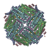
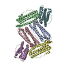
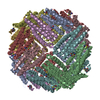
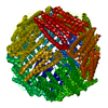
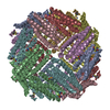
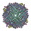
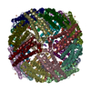
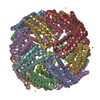
| Title | Crystal structure of the PmFTN variant E130A soaked in iron (overnight) | ||||||
 Components Components | Ferritin | ||||||
 Keywords Keywords | OXIDOREDUCTASE / ferritin / di-iron ferroxidase centre / 4-helix bundle | ||||||
| Function / homology |  Function and homology information Function and homology informationferroxidase / ferroxidase activity / ferric iron binding / iron ion transport / ferrous iron binding / intracellular iron ion homeostasis / identical protein binding / cytoplasm Similarity search - Function | ||||||
| Biological species |  Pseudo-nitzschia multiseries (Diatom) Pseudo-nitzschia multiseries (Diatom) | ||||||
| Method |  X-RAY DIFFRACTION / SYNCHROTRON / MOLECULAR REPLACEMENT / molecular replacement / Resolution: 2 Å X-RAY DIFFRACTION / SYNCHROTRON / MOLECULAR REPLACEMENT / molecular replacement / Resolution: 2 Å | ||||||
| Model details | overnight soaked | ||||||
 Authors Authors | Pfaffen, S. / Murphy, M.E.P. | ||||||
 Citation Citation | Journal: J.Biol.Chem. / Year: 2015 Title: A Diatom Ferritin Optimized for Iron Oxidation but Not Iron Storage. Authors: Pfaffen, S. / Bradley, J.M. / Abdulqadir, R. / Firme, M.R. / Moore, G.R. / Le Brun, N.E. / Murphy, M.E. | ||||||
| History |
|
- Structure visualization
| Structure viewer | Molecule: MolmilJmol/JSmol |
|---|
- Downloads & links
-Download
| PDBx/mmCIF format | 4zlw.cif.gz | 273.4 KB | Display | PDBx/mmCIF format |
|---|---|---|---|---|
| PDB format | pdb4zlw.ent.gz | 219.8 KB | Display | PDB format |
| PDBx/mmJSON format | 4zlw.json.gz | Tree view | PDBx/mmJSON format | |
| Others |  Other downloads Other downloads |
-Validation report
| Arichive directory | https://data.pdbj.org/pub/pdb/validation_reports/zl/4zlwftp://data.pdbj.org/pub/pdb/validation_reports/zl/4zlw | HTTPS FTP |
|---|
-Related structure data
| Related structure data |  4zkhSC 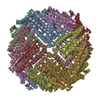 4zkwC  4zkxC  4zl5C  4zl6C  4zmcC S: Starting model for refinement C: citing same article ( |
|---|---|
| Similar structure data |


-Links
 PDBj
PDBj


- Assembly
| Deposited unit | 
| ||||||||||||||||||
|---|---|---|---|---|---|---|---|---|---|---|---|---|---|---|---|---|---|---|---|
| 1 | 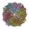
| ||||||||||||||||||
| Unit cell |
| ||||||||||||||||||
| Components on special symmetry positions |
|
-Components
| #1: Protein | Mass: 18808.945 Da / Num. of mol.: 8 / Fragment: iron storage protein / Mutation: E130A Source method: isolated from a genetically manipulated source Source: (gene. exp.) Pseudo-nitzschia multiseries (Diatom) / Gene: FTN / Plasmid: pET28 / Production host:  #2: Chemical | ChemComp-FE /   Mass: 55.845 Da / Num. of mol.: 37 / Source method: obtained synthetically / Formula: Fe Mass: 55.845 Da / Num. of mol.: 37 / Source method: obtained synthetically / Formula: Fe#3: Water | ChemComp-HOH / |  Mass: 18.015 Da / Num. of mol.: 510 / Source method: isolated from a natural source / Formula: H2O Mass: 18.015 Da / Num. of mol.: 510 / Source method: isolated from a natural source / Formula: H2O |
|---|
-Experimental details
-Experiment
| Experiment | Method: X-RAY DIFFRACTION / Number of used crystals: 1 |
|---|
- Sample preparation
| Crystal | Density Matthews: 2.99 Å3/Da / Density % sol: 58.92 % |
|---|---|
| Crystal grow | Temperature: 298 K / Method: vapor diffusion, hanging drop / Details: Sodiaum acetate pH 5.5, Ammonium sulfate, NaCl / Temp details: room temperature |
-Data collection
| Diffraction | Mean temperature: 100 K | ||||||||||||||||||||||||||||||||||||||||||||||||||||||||||||||||||||||||||||||||||||||||||||||||||||||||||||||
|---|---|---|---|---|---|---|---|---|---|---|---|---|---|---|---|---|---|---|---|---|---|---|---|---|---|---|---|---|---|---|---|---|---|---|---|---|---|---|---|---|---|---|---|---|---|---|---|---|---|---|---|---|---|---|---|---|---|---|---|---|---|---|---|---|---|---|---|---|---|---|---|---|---|---|---|---|---|---|---|---|---|---|---|---|---|---|---|---|---|---|---|---|---|---|---|---|---|---|---|---|---|---|---|---|---|---|---|---|---|---|---|
| Diffraction source | Source: SYNCHROTRON / Site: SSRL  / Beamline: BL7-1 / Wavelength: 1 Å / Beamline: BL7-1 / Wavelength: 1 Å | ||||||||||||||||||||||||||||||||||||||||||||||||||||||||||||||||||||||||||||||||||||||||||||||||||||||||||||||
| Detector | Type: ADSC QUANTUM 315r / Detector: CCD / Date: Jan 16, 2011 | ||||||||||||||||||||||||||||||||||||||||||||||||||||||||||||||||||||||||||||||||||||||||||||||||||||||||||||||
| Radiation | Protocol: SINGLE WAVELENGTH / Monochromatic (M) / Laue (L): M / Scattering type: x-ray | ||||||||||||||||||||||||||||||||||||||||||||||||||||||||||||||||||||||||||||||||||||||||||||||||||||||||||||||
| Radiation wavelength | Wavelength: 1 Å / Relative weight: 1 | ||||||||||||||||||||||||||||||||||||||||||||||||||||||||||||||||||||||||||||||||||||||||||||||||||||||||||||||
| Reflection | Resolution: 2→48.55 Å / Num. all: 120022 / Num. obs: 120022 / % possible obs: 100 % / Redundancy: 13.9 % / Rpim(I) all: 0.031 / Rrim(I) all: 0.115 / Rsym value: 0.107 / Net I/av σ(I): 5.63 / Net I/σ(I): 14.9 / Num. measured all: 1670804 | ||||||||||||||||||||||||||||||||||||||||||||||||||||||||||||||||||||||||||||||||||||||||||||||||||||||||||||||
| Reflection shell | Diffraction-ID: 1 / Rejects: _
|
-Phasing
| Phasing | Method: molecular replacement |
|---|
- Processing
| Software |
| |||||||||||||||||||||||||||||||||||||||||||||||||||||||||||||||||||||||||||
|---|---|---|---|---|---|---|---|---|---|---|---|---|---|---|---|---|---|---|---|---|---|---|---|---|---|---|---|---|---|---|---|---|---|---|---|---|---|---|---|---|---|---|---|---|---|---|---|---|---|---|---|---|---|---|---|---|---|---|---|---|---|---|---|---|---|---|---|---|---|---|---|---|---|---|---|---|
| Refinement | Method to determine structure: MOLECULAR REPLACEMENT Starting model: 4ZKH Resolution: 2→48.55 Å / Cor.coef. Fo:Fc: 0.954 / Cor.coef. Fo:Fc free: 0.939 / WRfactor Rfree: 0.2181 / WRfactor Rwork: 0.1787 / FOM work R set: 0.8407 / SU B: 3.978 / SU ML: 0.108 / SU R Cruickshank DPI: 0.1457 / SU Rfree: 0.1409 / Cross valid method: THROUGHOUT / σ(F): 0 / ESU R: 0.146 / ESU R Free: 0.141 / Stereochemistry target values: MAXIMUM LIKELIHOOD Details: HYDROGENS HAVE BEEN ADDED IN THE RIDING POSITIONS U VALUES : REFINED INDIVIDUALLY
| |||||||||||||||||||||||||||||||||||||||||||||||||||||||||||||||||||||||||||
| Solvent computation | Ion probe radii: 0.8 Å / Shrinkage radii: 0.8 Å / VDW probe radii: 1.2 Å / Solvent model: MASK | |||||||||||||||||||||||||||||||||||||||||||||||||||||||||||||||||||||||||||
| Displacement parameters | Biso max: 117.9 Å2 / Biso mean: 40.874 Å2 / Biso min: 17.2 Å2
| |||||||||||||||||||||||||||||||||||||||||||||||||||||||||||||||||||||||||||
| Refinement step | Cycle: final / Resolution: 2→48.55 Å
| |||||||||||||||||||||||||||||||||||||||||||||||||||||||||||||||||||||||||||
| Refine LS restraints |
| |||||||||||||||||||||||||||||||||||||||||||||||||||||||||||||||||||||||||||
| LS refinement shell | Resolution: 2→2.052 Å / Total num. of bins used: 20
|
