 Movie
Movie Controller

[English] 日本語
 Yorodumi
- PDB-4toe: 2.20A resolution structure of Iron Bound BfrB (D34F) from Pseudom... -
+ Open data

- Basic information
| Entry | Database: PDB / ID: 4toe | ||||||
|---|---|---|---|---|---|---|---|
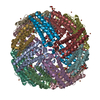
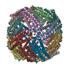
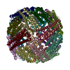
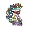
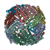
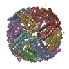
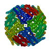
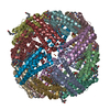
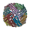
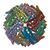
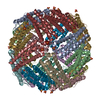
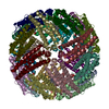
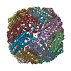
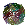
| Title | 2.20A resolution structure of Iron Bound BfrB (D34F) from Pseudomonas aeruginosa | ||||||
 Components Components | Bacterioferritin | ||||||
 Keywords Keywords | OXIDOREDUCTASE / ELECTRON TRANSPORT / IRON STORAGE / iron binding / iron mobilization | ||||||
| Function / homology |  Function and homology information Function and homology informationiron ion sequestering activity / ferritin complex / ferroxidase / ferroxidase activity / ferric iron binding / iron ion transport / intracellular iron ion homeostasis / iron ion binding / heme binding / cytosol Similarity search - Function | ||||||
| Biological species |   Pseudomonas aeruginosa (bacteria) Pseudomonas aeruginosa (bacteria) | ||||||
| Method |  X-RAY DIFFRACTION / SYNCHROTRON / MOLECULAR REPLACEMENT / molecular replacement / Resolution: 2.2 Å X-RAY DIFFRACTION / SYNCHROTRON / MOLECULAR REPLACEMENT / molecular replacement / Resolution: 2.2 Å | ||||||
 Authors Authors | Lovell, S. / Battaile, K.P. / Yao, H. / Kumar, R. / Eshelman, K. / Rivera, M. | ||||||
| Funding support |  United States, 1items United States, 1items
| ||||||
 Citation Citation | Journal: Biochemistry / Year: 2015 Title: Concerted motions networking pores and distant ferroxidase centers enable bacterioferritin function and iron traffic. Authors: Yao, H. / Rui, H. / Kumar, R. / Eshelman, K. / Lovell, S. / Battaile, K.P. / Im, W. / Rivera, M. | ||||||
| History |
|
- Structure visualization
| Structure viewer | Molecule: MolmilJmol/JSmol |
|---|
- Downloads & links
-Download
| PDBx/mmCIF format | 4toe.cif.gz | 827.2 KB | Display | PDBx/mmCIF format |
|---|---|---|---|---|
| PDB format | pdb4toe.ent.gz | 682.1 KB | Display | PDB format |
| PDBx/mmJSON format | 4toe.json.gz | Tree view | PDBx/mmJSON format | |
| Others |  Other downloads Other downloads |
-Validation report
| Arichive directory | https://data.pdbj.org/pub/pdb/validation_reports/to/4toeftp://data.pdbj.org/pub/pdb/validation_reports/to/4toe | HTTPS FTP |
|---|
-Related structure data
| Related structure data |  4to9C  4toaC  4tobC  4tocC 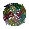 4todC 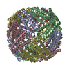 4tofC 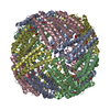 4togC  4tohC 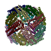 3is7S C: citing same article ( S: Starting model for refinement |
|---|---|
| Similar structure data |


-Links
 PDBj
PDBj


- Assembly
| Deposited unit | 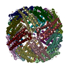
| ||||||||
|---|---|---|---|---|---|---|---|---|---|
| 1 |
| ||||||||
| Unit cell |
| ||||||||
| Details | The asymmetric unit contains one biological unit |
-Components
-Protein , 1 types, 24 molecules ABCDEFGHIJKLMNOPQRSTUVWX
| #1: Protein | Mass: 18612.254 Da / Num. of mol.: 24 / Mutation: D34F Source method: isolated from a genetically manipulated source Source: (gene. exp.) Pseudomonas aeruginosa (bacteria) / Strain: ATCC 15692 / PAO1 / 1C / PRS 101 / LMG 12228 / Gene: bfrB, PA3531 / Plasmid: pET11a / Production host: |
|---|
-Non-polymers , 5 types, 2448 molecules 


| #2: Chemical | ChemComp-FE2 /  Mass: 55.845 Da / Num. of mol.: 144 / Source method: obtained synthetically / Formula: Fe Mass: 55.845 Da / Num. of mol.: 144 / Source method: obtained synthetically / Formula: Fe#3: Chemical | ChemComp-HEM /  Mass: 616.487 Da / Num. of mol.: 12 / Source method: obtained synthetically / Formula: C34H32FeN4O4 Mass: 616.487 Da / Num. of mol.: 12 / Source method: obtained synthetically / Formula: C34H32FeN4O4#4: Chemical | ChemComp-K /  Mass: 39.098 Da / Num. of mol.: 6 / Source method: obtained synthetically / Formula: K Mass: 39.098 Da / Num. of mol.: 6 / Source method: obtained synthetically / Formula: K#5: Chemical | ChemComp-SO4 /  Mass: 96.063 Da / Num. of mol.: 8 / Source method: obtained synthetically / Formula: SO4 Mass: 96.063 Da / Num. of mol.: 8 / Source method: obtained synthetically / Formula: SO4#6: Water | ChemComp-HOH / | Mass: 18.015 Da / Num. of mol.: 2278 / Source method: isolated from a natural source / Formula: H2O |
|---|
-Experimental details
-Experiment
| Experiment | Method: X-RAY DIFFRACTION / Number of used crystals: 1 |
|---|
- Sample preparation
| Crystal | Colour: Red Prism / Density Matthews: 2.96 Å3/Da / Density % sol: 58.46 % Description: THE STRUCTURE FACTOR FILE CONTAINS FRIEDEL PAIRS |
|---|---|
| Crystal grow | Temperature: 293 K / Method: vapor diffusion / pH: 6 / Details: 35% (v/v) MPD, 0.1M MES, 0.2M Lithium Sulfate |
-Data collection
| Diffraction | Mean temperature: 100 K | |||||||||||||||||||||||||||
|---|---|---|---|---|---|---|---|---|---|---|---|---|---|---|---|---|---|---|---|---|---|---|---|---|---|---|---|---|
| Diffraction source | Source: SYNCHROTRON / Site: APS / Beamline: 17-ID / Wavelength: 1.73769 Å | |||||||||||||||||||||||||||
| Detector | Type: DECTRIS PILATUS 6M / Detector: PIXEL / Date: Nov 12, 2011 | |||||||||||||||||||||||||||
| Radiation | Protocol: SINGLE WAVELENGTH / Monochromatic (M) / Laue (L): M / Scattering type: x-ray | |||||||||||||||||||||||||||
| Radiation wavelength | Wavelength: 1.73769 Å / Relative weight: 1 | |||||||||||||||||||||||||||
| Reflection | Resolution: 2.2→49.39 Å / Num. obs: 257692 / % possible obs: 96.3 % / Redundancy: 5.8 % / Biso Wilson estimate: 21.55 Å2 / CC1/2: 0.998 / Rmerge(I) obs: 0.078 / Rpim(I) all: 0.034 / Net I/σ(I): 18.1 / Num. measured all: 1490791 | |||||||||||||||||||||||||||
| Reflection shell | Diffraction-ID: 1 / Rejects: _
|
-Phasing
| Phasing | Method: molecular replacement |
|---|
- Processing
| Software |
| |||||||||||||||||||||||||||||||||||||||||||||||||||||||||||||||||||||||||||||||||||||||||||||||||||||||||||||||||||||||||||||||||||||||||||||||||||||||||||||||||||||||||||||||||||||||||||||||||||||||||||||||||||||||||
|---|---|---|---|---|---|---|---|---|---|---|---|---|---|---|---|---|---|---|---|---|---|---|---|---|---|---|---|---|---|---|---|---|---|---|---|---|---|---|---|---|---|---|---|---|---|---|---|---|---|---|---|---|---|---|---|---|---|---|---|---|---|---|---|---|---|---|---|---|---|---|---|---|---|---|---|---|---|---|---|---|---|---|---|---|---|---|---|---|---|---|---|---|---|---|---|---|---|---|---|---|---|---|---|---|---|---|---|---|---|---|---|---|---|---|---|---|---|---|---|---|---|---|---|---|---|---|---|---|---|---|---|---|---|---|---|---|---|---|---|---|---|---|---|---|---|---|---|---|---|---|---|---|---|---|---|---|---|---|---|---|---|---|---|---|---|---|---|---|---|---|---|---|---|---|---|---|---|---|---|---|---|---|---|---|---|---|---|---|---|---|---|---|---|---|---|---|---|---|---|---|---|---|---|---|---|---|---|---|---|---|---|---|---|---|---|---|---|---|
| Refinement | Method to determine structure: MOLECULAR REPLACEMENT Starting model: 3IS7 Resolution: 2.2→48.39 Å / SU ML: 0.19 / Cross valid method: FREE R-VALUE / σ(F): 1.92 / Phase error: 18.11 / Stereochemistry target values: ML / Details: THE FRIEDEL PAIRS WERE USED IN REFINEMENT
| |||||||||||||||||||||||||||||||||||||||||||||||||||||||||||||||||||||||||||||||||||||||||||||||||||||||||||||||||||||||||||||||||||||||||||||||||||||||||||||||||||||||||||||||||||||||||||||||||||||||||||||||||||||||||
| Solvent computation | Shrinkage radii: 0.9 Å / VDW probe radii: 1.11 Å / Solvent model: FLAT BULK SOLVENT MODEL | |||||||||||||||||||||||||||||||||||||||||||||||||||||||||||||||||||||||||||||||||||||||||||||||||||||||||||||||||||||||||||||||||||||||||||||||||||||||||||||||||||||||||||||||||||||||||||||||||||||||||||||||||||||||||
| Displacement parameters | Biso max: 80.48 Å2 / Biso mean: 24.7504 Å2 / Biso min: 10.77 Å2 | |||||||||||||||||||||||||||||||||||||||||||||||||||||||||||||||||||||||||||||||||||||||||||||||||||||||||||||||||||||||||||||||||||||||||||||||||||||||||||||||||||||||||||||||||||||||||||||||||||||||||||||||||||||||||
| Refinement step | Cycle: final / Resolution: 2.2→48.39 Å
| |||||||||||||||||||||||||||||||||||||||||||||||||||||||||||||||||||||||||||||||||||||||||||||||||||||||||||||||||||||||||||||||||||||||||||||||||||||||||||||||||||||||||||||||||||||||||||||||||||||||||||||||||||||||||
| Refine LS restraints |
| |||||||||||||||||||||||||||||||||||||||||||||||||||||||||||||||||||||||||||||||||||||||||||||||||||||||||||||||||||||||||||||||||||||||||||||||||||||||||||||||||||||||||||||||||||||||||||||||||||||||||||||||||||||||||
| LS refinement shell | Refine-ID: X-RAY DIFFRACTION / Total num. of bins used: 30
|
