Mass: 18.015 Da / Num. of mol.: 282 / Source method: isolated from a natural source / Formula: H2O
Sequence details
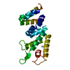
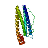
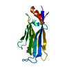
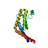
ACCORDING TO THE AUTHORS, THE PROTEIN WAS DERIVED FROM OAS TG'S LAMBDA REPRESSOR PROTEIN EXPRESSION ...ACCORDING TO THE AUTHORS, THE PROTEIN WAS DERIVED FROM OAS TG'S LAMBDA REPRESSOR PROTEIN EXPRESSION VECTOR WHICH CARRIES THE CHANGE OF THE FIRST N-TERM AND THE LAST C-TERM RESIDUES. THESE TWO CHANGES CAME FROM THE PREPARATION OF THE EXPRESSION SYSTEM, POSSIBLY THE RESTRICTION ENZYME DIGESTION SITE DESIGN WHEN THE DNA SEQUENCE WAS CUT FROM THE WILD-TYPE PROTEIN AND INSERTED INTO THE VECTOR
-
Experimental details
-
Experiment
Experiment
Method: X-RAY DIFFRACTION / Number of used crystals: 1
-
Sample preparation
Crystal
Density Matthews: 2.3 Å3/Da / Density % sol: 46.59 %
Crystal grow
Temperature: 277 K / Method: vapor diffusion, hanging drop / pH: 6.5 Details: PEG, pH 6.5, VAPOR DIFFUSION, HANGING DROP, temperature 277K
Type: MAR CCD 130 mm / Detector: CCD / Date: Sep 26, 2008
Radiation
Protocol: SINGLE WAVELENGTH / Monochromatic (M) / Laue (L): M / Scattering type: x-ray
Radiation wavelength
Wavelength: 0.97 Å / Relative weight: 1
Reflection
Resolution: 1.64→30 Å / Num. obs: 19544 / % possible obs: 99.1 % / Redundancy: 7.3 % / Biso Wilson estimate: 23.52 Å2 / Rmerge(I) obs: 0.067
Reflection shell
Resolution: 1.64→1.7 Å / Redundancy: 6.6 % / % possible all: 90.9
-
Processing
Software
Name
Classification
MAR345dtb
datacollection
AMoRE
phasing
SHELXL-97
refinement
HKL-2000
datareduction
SCALEPACK
datascaling
Refinement
Method to determine structure: AB INITIO / Resolution: 1.64→10 Å / Cross valid method: FREE R / σ(F): 0 / Stereochemistry target values: ENGH AND HUBER Details: ANISOTROPIC SCALING APPLIED BY THE METHOD OF PARKIN, MOEZZI & HOPE
Rfactor
Num. reflection
% reflection
Selection details
Rwork
0.1879
-
-
-
obs
0.1889
17329
93.8 %
-
all
-
18248
-
-
Rfree
-
-
-
RANDOM
Refinement step
Cycle: LAST / Resolution: 1.64→10 Å
Protein
Nucleic acid
Ligand
Solvent
Total
Num. atoms
1236
0
0
282
1518
Refine LS restraints
Refine-ID
Type
Dev ideal
X-RAY DIFFRACTION
s_bond_d
0.007
X-RAY DIFFRACTION
s_angle_d
1.957
X-RAY DIFFRACTION
s_similar_dist
0
X-RAY DIFFRACTION
s_from_restr_planes
0.022
X-RAY DIFFRACTION
s_zero_chiral_vol
0.036
X-RAY DIFFRACTION
s_non_zero_chiral_vol
0.043
X-RAY DIFFRACTION
s_anti_bump_dis_restr
0.017
X-RAY DIFFRACTION
s_rigid_bond_adp_cmpnt
0
X-RAY DIFFRACTION
s_similar_adp_cmpnt
0.07
X-RAY DIFFRACTION
s_approx_iso_adps
0
+
About Yorodumi
-
News
-
Feb 9, 2022. New format data for meta-information of EMDB entries
New format data for meta-information of EMDB entries
Version 3 of the EMDB header file is now the official format.
The previous official version 1.9 will be removed from the archive.
In the structure databanks used in Yorodumi, some data are registered as the other names, "COVID-19 virus" and "2019-nCoV". Here are the details of the virus and the list of structure data.
Jan 31, 2019. EMDB accession codes are about to change! (news from PDBe EMDB page)
EMDB accession codes are about to change! (news from PDBe EMDB page)
The allocation of 4 digits for EMDB accession codes will soon come to an end. Whilst these codes will remain in use, new EMDB accession codes will include an additional digit and will expand incrementally as the available range of codes is exhausted. The current 4-digit format prefixed with “EMD-” (i.e. EMD-XXXX) will advance to a 5-digit format (i.e. EMD-XXXXX), and so on. It is currently estimated that the 4-digit codes will be depleted around Spring 2019, at which point the 5-digit format will come into force.
The EM Navigator/Yorodumi systems omit the EMD- prefix.
Related info.:Q: What is EMD? / ID/Accession-code notation in Yorodumi/EM Navigator
Yorodumi is a browser for structure data from EMDB, PDB, SASBDB, etc.
This page is also the successor to EM Navigator detail page, and also detail information page/front-end page for Omokage search.
The word "yorodu" (or yorozu) is an old Japanese word meaning "ten thousand". "mi" (miru) is to see.
Related info.:EMDB / PDB / SASBDB / Comparison of 3 databanks / Yorodumi Search / Aug 31, 2016. New EM Navigator & Yorodumi / Yorodumi Papers / Jmol/JSmol / Function and homology information / Changes in new EM Navigator and Yorodumi
 Movie
Movie Controller


 Open data
 Basic information
 Components
 Keywords
 Function and homology information
 Enterobacteria phage lambda (virus)
 X-RAY DIFFRACTION /
  Authors
 Citation
 Structure visualization
 Downloads & links
 Other downloads


 PDBj
PDBj


 Assembly


 Mass: 18.015 Da / Num. of mol.: 282 / Source method: isolated from a natural source / Formula: H2O
 Sample preparation
 / Beamline: BL9-3 / Wavelength: 0.97 Å
 Processing