 ムービー
ムービー コントローラー


+ データを開く

- 基本情報
基本情報
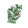
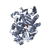
| 登録情報 | データベース: PDB / ID: 1kdw | ||||||
|---|---|---|---|---|---|---|---|
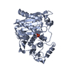
| タイトル | X-ray crystal structure of AmpC beta-lactamase from E. coli in complex with the inhibitor 4-carboxyphenylboronic acid | ||||||
 要素 要素 | beta-lactamase | ||||||
 キーワード キーワード | HYDROLASE / cephalosporinase / beta-lactamase / serine hydrolase / phenylboronic acid inhibitor | ||||||
| 機能・相同性 |  機能・相同性情報 機能・相同性情報antibiotic catabolic process / beta-lactamase activity / beta-lactamase / outer membrane-bounded periplasmic space / response to antibiotic 類似検索 - 分子機能 | ||||||
| 生物種 |  | ||||||
| 手法 |  X線回折 / 分子置換 / 解像度: 2.28 Å X線回折 / 分子置換 / 解像度: 2.28 Å | ||||||
 データ登録者 データ登録者 | Powers, R.A. / Shoichet, B.K. | ||||||
 引用 引用 | ジャーナル: J.Med.Chem. / 年: 2002 タイトル: Structure-based approach for binding site identification on AmpC beta-lactamase. 著者: Powers, R.A. / Shoichet, B.K. | ||||||
| 履歴 |
|
- 構造の表示
構造の表示
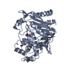
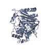
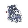
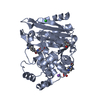
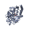
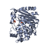
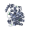
| 構造ビューア | 分子: MolmilJmol/JSmol |
|---|
- ダウンロードとリンク
-ダウンロード
| PDBx/mmCIF形式 | 1kdw.cif.gz | 152.9 KB | 表示 | PDBx/mmCIF形式 |
|---|---|---|---|---|
| PDB形式 | pdb1kdw.ent.gz | 120.3 KB | 表示 | PDB形式 |
| PDBx/mmJSON形式 | 1kdw.json.gz | ツリー表示 | PDBx/mmJSON形式 | |
| その他 |  その他のダウンロード その他のダウンロード |
-検証レポート
| アーカイブディレクトリ | https://data.pdbj.org/pub/pdb/validation_reports/kd/1kdwftp://data.pdbj.org/pub/pdb/validation_reports/kd/1kdw | HTTPS FTP |
|---|
-関連構造データ


-リンク
 PDBj
PDBj
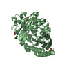
- 集合体
集合体
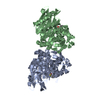
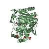
| 登録構造単位 | 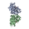
| ||||||||
|---|---|---|---|---|---|---|---|---|---|
| 1 | 
| ||||||||
| 2 | 
| ||||||||
| 単位格子 |
|
-要素
| #1: タンパク質 | 分子量: 39587.922 Da / 分子数: 2 / 由来タイプ: 組換発現 / 由来: (組換発現) #2: 化合物 | ChemComp-PO4 / |   分子量: 94.971 Da / 分子数: 1 / 由来タイプ: 合成 / 式: PO4 分子量: 94.971 Da / 分子数: 1 / 由来タイプ: 合成 / 式: PO4#3: 化合物 | 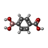  分子量: 165.939 Da / 分子数: 2 / 由来タイプ: 合成 / 式: C7H7BO4 分子量: 165.939 Da / 分子数: 2 / 由来タイプ: 合成 / 式: C7H7BO4#4: 水 | ChemComp-HOH / |  分子量: 18.015 Da / 分子数: 263 / 由来タイプ: 天然 / 式: H2O 分子量: 18.015 Da / 分子数: 263 / 由来タイプ: 天然 / 式: H2OHas protein modification | Y | |
|---|
-実験情報
-実験
| 実験 | 手法: X線回折 / 使用した結晶の数: 1 |
|---|
- 試料調製
試料調製
| 結晶 | マシュー密度: 2.55 Å3/Da / 溶媒含有率: 51.71 % | ||||||||||||||||||||||||
|---|---|---|---|---|---|---|---|---|---|---|---|---|---|---|---|---|---|---|---|---|---|---|---|---|---|
| 結晶化 | 温度: 296 K / 手法: 蒸気拡散法, ハンギングドロップ法 / pH: 8.7 詳細: 1.7 M potassium phosphate, 485 uM 4-carboxyphenylboronic acid, pH 8.7, VAPOR DIFFUSION, HANGING DROP, temperature 296K | ||||||||||||||||||||||||
| 結晶化 | *PLUS 温度: 23 ℃ / 詳細: used microseeding | ||||||||||||||||||||||||
| 溶液の組成 | *PLUS
|
-データ収集
| 回折 | 平均測定温度: 103 K |
|---|---|
| 放射光源 | 由来: 回転陽極 / タイプ: RIGAKU RU200 / 波長: 1.5418 Å |
| 検出器 | タイプ: RIGAKU RAXIS IIC / 検出器: IMAGE PLATE / 日付: 1998年10月1日 |
| 放射 | プロトコル: SINGLE WAVELENGTH / 単色(M)・ラウエ(L): M / 散乱光タイプ: x-ray |
| 放射波長 | 波長: 1.5418 Å / 相対比: 1 |
| 反射 | 解像度: 2.28→20 Å / Num. all: 36794 / Num. obs: 33450 / % possible obs: 91 % / Observed criterion σ(I): -3 / 冗長度: 2.7 % / Biso Wilson estimate: 25.46 Å2 / Rmerge(I) obs: 0.075 / Net I/σ(I): 16.2 |
| 反射 シェル | 解像度: 2.28→2.37 Å / Rmerge(I) obs: 0.19 / Mean I/σ(I) obs: 3.5 / % possible all: 62.6 |
| 反射 | *PLUS 最低解像度: 20 Å / Num. measured all: 90630 / Rmerge(I) obs: 0.075 |
| 反射 シェル | *PLUS % possible obs: 62.6 % / Rmerge(I) obs: 0.19 |
- 解析
解析
| ソフトウェア |
| |||||||||||||||||||||||||
|---|---|---|---|---|---|---|---|---|---|---|---|---|---|---|---|---|---|---|---|---|---|---|---|---|---|---|
| 精密化 | 構造決定の手法: 分子置換 開始モデル: PDB entry 1C3B 解像度: 2.28→20 Å / σ(F): 2 / 立体化学のターゲット値: Engh & Huber
| |||||||||||||||||||||||||
| Refine analyze |
| |||||||||||||||||||||||||
| 精密化ステップ | サイクル: LAST / 解像度: 2.28→20 Å
| |||||||||||||||||||||||||
| 拘束条件 |
| |||||||||||||||||||||||||
| LS精密化 シェル | 解像度: 2.28→2.37 Å
| |||||||||||||||||||||||||
| 精密化 | *PLUS 最低解像度: 20 Å / % reflection Rfree: 10 % / Rfactor Rfree: 0.234 / Rfactor Rwork: 0.186 | |||||||||||||||||||||||||
| 溶媒の処理 | *PLUS | |||||||||||||||||||||||||
| 原子変位パラメータ | *PLUS | |||||||||||||||||||||||||
| LS精密化 シェル | *PLUS Rfactor Rfree: 0.345 / Rfactor Rwork: 0.252 |
