 Movie
Movie Controller


+ Open data

- Basic information
| Entry | Database: PDB / ID: 1k61 | ||||||
|---|---|---|---|---|---|---|---|
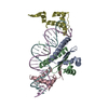
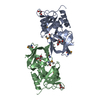
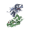
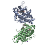
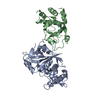
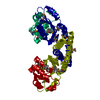
| Title | MATALPHA2 HOMEODOMAIN BOUND TO DNA | ||||||
 Components Components |
| ||||||
 Keywords Keywords | TRANSCRIPTION/DNA / PROTEIN-DNA COMPLEX / HOMEODOMAIN / HOOGSTEEN BASE PAIR / TRANSCRIPTION-DNA COMPLEX | ||||||
| Function / homology |  Function and homology information Function and homology information: / regulation of mating-type specific transcription, DNA-templated / RNA polymerase II transcription repressor complex / DNA binding, bending / DNA-binding transcription repressor activity, RNA polymerase II-specific / sequence-specific DNA binding / negative regulation of transcription by RNA polymerase II / nucleus Similarity search - Function | ||||||
| Method |  X-RAY DIFFRACTION / MIR, molecular replacement / Resolution: 2.1 Å X-RAY DIFFRACTION / MIR, molecular replacement / Resolution: 2.1 Å | ||||||
 Authors Authors | Aishima, J. / Gitti, R.K. / Noah, J.E. / Gan, H.H. / Schlick, T. / Wolberger, C. | ||||||
 Citation Citation | Journal: NUCLEIC ACIDS RES. / Year: 2002 Title: A Hoogsteen base pair embedded in undistorted B-DNA Authors: Aishima, J. / Gitti, R.K. / Noah, J.E. / Gan, H.H. / Schlick, T. / Wolberger, C. | ||||||
| History |
|
- Structure visualization
| Structure viewer | Molecule: MolmilJmol/JSmol |
|---|
- Downloads & links
-Download
| PDBx/mmCIF format | 1k61.cif.gz | 89 KB | Display | PDBx/mmCIF format |
|---|---|---|---|---|
| PDB format | pdb1k61.ent.gz | 63.3 KB | Display | PDB format |
| PDBx/mmJSON format | 1k61.json.gz | Tree view | PDBx/mmJSON format | |
| Others |  Other downloads Other downloads |
-Validation report
| Summary document | 1k61_validation.pdf.gz | 468.5 KB | Display | wwPDB validaton report |
|---|---|---|---|---|
| Full document | 1k61_full_validation.pdf.gz | 474.1 KB | Display | |
| Data in XML | 1k61_validation.xml.gz | 14.6 KB | Display | |
| Data in CIF | 1k61_validation.cif.gz | 20.9 KB | Display | |
| Arichive directory | https://data.pdbj.org/pub/pdb/validation_reports/k6/1k61ftp://data.pdbj.org/pub/pdb/validation_reports/k6/1k61 | HTTPS FTP |
-Related structure data
| Related structure data |  1aplS S: Starting model for refinement |
|---|---|
| Similar structure data |


-Links
 PDBj
PDBj


- Assembly
| Deposited unit | 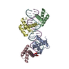
| ||||||||
|---|---|---|---|---|---|---|---|---|---|
| 1 |
| ||||||||
| Unit cell |
|
-Components
| #1: DNA chain | Mass: 6381.160 Da / Num. of mol.: 1 / Source method: obtained synthetically Details: This sequence is derived from the STE6 promoter region, with the MCM1 binding sites removed. | ||
|---|---|---|---|
| #2: DNA chain | Mass: 6613.103 Da / Num. of mol.: 1 / Source method: obtained synthetically Details: This sequence is derived from the STE6 promoter region, with the MCM1 binding sites removed. | ||
| #3: Protein | Mass: 7275.384 Da / Num. of mol.: 4 / Fragment: RESIDUES 132-191, HOMEODOMAIN / Source method: obtained synthetically Details: The sequence naturally occurs in yeast. The protein was synthesized by the FMOC method. References: UniProt: P01367, UniProt: P0CY08*PLUS #4: Water | ChemComp-HOH / |  Mass: 18.015 Da / Num. of mol.: 195 / Source method: isolated from a natural source / Formula: H2O Mass: 18.015 Da / Num. of mol.: 195 / Source method: isolated from a natural source / Formula: H2O |
-Experimental details
-Experiment
| Experiment | Method: X-RAY DIFFRACTION / Number of used crystals: 1 |
|---|
- Sample preparation
| Crystal | Density Matthews: 2.09 Å3/Da / Density % sol: 41.2 % | ||||||||||||
|---|---|---|---|---|---|---|---|---|---|---|---|---|---|
| Crystal grow | Temperature: 291 K / Method: vapor diffusion, hanging drop / pH: 9 Details: PEG 6000, Bicine, pH 9.0, VAPOR DIFFUSION, HANGING DROP, temperature 291K | ||||||||||||
| Components of the solutions |
|
-Data collection
| Diffraction | Mean temperature: 100 K |
|---|---|
| Diffraction source | Source: ROTATING ANODE / Type: RIGAKU RU200 / Wavelength: 1.5418 Å |
| Detector | Type: RIGAKU RAXIS IV / Detector: IMAGE PLATE / Date: Jun 24, 1998 / Details: Yale mirrors |
| Radiation | Monochromator: YALE MIRRORS / Protocol: SINGLE WAVELENGTH / Monochromatic (M) / Laue (L): M / Scattering type: x-ray |
| Radiation wavelength | Wavelength: 1.5418 Å / Relative weight: 1 |
| Reflection | Resolution: 2→16 Å / Num. all: 23364 / Num. obs: 23014 / % possible obs: 98.5 % / Observed criterion σ(F): 0 / Observed criterion σ(I): 0 / Redundancy: 2.07 % / Biso Wilson estimate: 33.8 Å2 / Rmerge(I) obs: 0.048 / Net I/σ(I): 14 |
| Reflection shell | Resolution: 2→2.07 Å / Redundancy: 2.3 % / Rmerge(I) obs: 0.287 / Mean I/σ(I) obs: 2.5 / Num. unique all: 2897 / % possible all: 81 |
| Reflection | *PLUS Lowest resolution: 16 Å |
- Processing
| Software |
| ||||||||||||||||||||||||||||||||||||||||||||||||||||||||||||||||||||||||||||||||
|---|---|---|---|---|---|---|---|---|---|---|---|---|---|---|---|---|---|---|---|---|---|---|---|---|---|---|---|---|---|---|---|---|---|---|---|---|---|---|---|---|---|---|---|---|---|---|---|---|---|---|---|---|---|---|---|---|---|---|---|---|---|---|---|---|---|---|---|---|---|---|---|---|---|---|---|---|---|---|---|---|---|
| Refinement | Method to determine structure: MIR, molecular replacement Starting model: PDB ENTRY 1APL Resolution: 2.1→16.01 Å / Rfactor Rfree error: 0.006 / Data cutoff high absF: 23852424.02 / Data cutoff low absF: 0 / Isotropic thermal model: RESTRAINED / Cross valid method: THROUGHOUT / σ(F): 0 Details: Refinement Target values for the DNA as described in: G.Parkinson, J.Vojtechovsky, L.Clowney, A.T.Brunger, H.M.Berman, New Parameters for the Refinement of Nucleic Acid Containing ...Details: Refinement Target values for the DNA as described in: G.Parkinson, J.Vojtechovsky, L.Clowney, A.T.Brunger, H.M.Berman, New Parameters for the Refinement of Nucleic Acid Containing Structures, Acta Cryst. D, 52, 57-64 (1996). Modified for 5-iodouracil residue.
| ||||||||||||||||||||||||||||||||||||||||||||||||||||||||||||||||||||||||||||||||
| Solvent computation | Solvent model: FLAT MODEL / Bsol: 44.8193 Å2 / ksol: 0.327842 e/Å3 | ||||||||||||||||||||||||||||||||||||||||||||||||||||||||||||||||||||||||||||||||
| Displacement parameters | Biso mean: 35.7 Å2
| ||||||||||||||||||||||||||||||||||||||||||||||||||||||||||||||||||||||||||||||||
| Refine analyze |
| ||||||||||||||||||||||||||||||||||||||||||||||||||||||||||||||||||||||||||||||||
| Refinement step | Cycle: LAST / Resolution: 2.1→16.01 Å
| ||||||||||||||||||||||||||||||||||||||||||||||||||||||||||||||||||||||||||||||||
| Refine LS restraints |
| ||||||||||||||||||||||||||||||||||||||||||||||||||||||||||||||||||||||||||||||||
| LS refinement shell | Resolution: 2.1→2.23 Å / Rfactor Rfree error: 0.02 / Total num. of bins used: 6
| ||||||||||||||||||||||||||||||||||||||||||||||||||||||||||||||||||||||||||||||||
| Xplor file |
| ||||||||||||||||||||||||||||||||||||||||||||||||||||||||||||||||||||||||||||||||
| Refinement | *PLUS Highest resolution: 2.1 Å / Rfactor Rfree: 0.278 / Rfactor Rwork: 0.222 | ||||||||||||||||||||||||||||||||||||||||||||||||||||||||||||||||||||||||||||||||
| Solvent computation | *PLUS | ||||||||||||||||||||||||||||||||||||||||||||||||||||||||||||||||||||||||||||||||
| Displacement parameters | *PLUS | ||||||||||||||||||||||||||||||||||||||||||||||||||||||||||||||||||||||||||||||||
| Refine LS restraints | *PLUS
|
