 Movie
Movie Controller

[English] 日本語
 Yorodumi
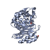
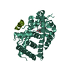
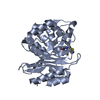
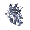
- PDB-3ry9: Crystal Structure of the Resurrected Ancestral Glucocorticoid Rec... -
+ Open data

- Basic information
| Entry | Database: PDB / ID: 3ry9 | ||||||
|---|---|---|---|---|---|---|---|
| Title | Crystal Structure of the Resurrected Ancestral Glucocorticoid Receptor 1 in complex with DOC | ||||||
 Components Components | Ancestral Glucocorticoid Receptor 1 | ||||||
 Keywords Keywords | STEROID BINDING PROTEIN / resurrected protein / steroid receptor / nuclear receptor / common ancestor / evolution | ||||||
| Function / homology | Retinoid X Receptor / Retinoid X Receptor / Orthogonal Bundle / Mainly Alpha / DESOXYCORTICOSTERONE Function and homology information Function and homology information | ||||||
| Biological species | artificial gene (others) | ||||||
| Method |  X-RAY DIFFRACTION / SYNCHROTRON / MOLECULAR REPLACEMENT / Resolution: 1.95 Å X-RAY DIFFRACTION / SYNCHROTRON / MOLECULAR REPLACEMENT / Resolution: 1.95 Å | ||||||
 Authors Authors | Ortlund, E.A. | ||||||
 Citation Citation | Journal: PLoS Genet / Year: 2011 Title: Mechanisms for the evolution of a derived function in the ancestral glucocorticoid receptor. Authors: Carroll, S.M. / Ortlund, E.A. / Thornton, J.W. | ||||||
| History |
|
- Structure visualization
| Structure viewer | Molecule: MolmilJmol/JSmol |
|---|
- Downloads & links
-Download
| PDBx/mmCIF format | 3ry9.cif.gz | 216.4 KB | Display | PDBx/mmCIF format |
|---|---|---|---|---|
| PDB format | pdb3ry9.ent.gz | 173.6 KB | Display | PDB format |
| PDBx/mmJSON format | 3ry9.json.gz | Tree view | PDBx/mmJSON format | |
| Others |  Other downloads Other downloads |
-Validation report
| Arichive directory | https://data.pdbj.org/pub/pdb/validation_reports/ry/3ry9ftp://data.pdbj.org/pub/pdb/validation_reports/ry/3ry9 | HTTPS FTP |
|---|
-Related structure data
| Related structure data |  2q3yS S: Starting model for refinement |
|---|---|
| Similar structure data |


-Links
 PDBj
PDBj- Assembly
| Deposited unit | 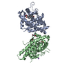
| ||||||||
|---|---|---|---|---|---|---|---|---|---|
| 1 | 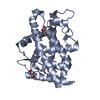
| ||||||||
| 2 | 
| ||||||||
| Unit cell |
|
-Components
| #1: Protein | Mass: 28797.551 Da / Num. of mol.: 2 / Fragment: ligand binding domain Source method: isolated from a genetically manipulated source Source: (gene. exp.) artificial gene (others) / Gene: resurrected gene / Plasmid: pLIC_MBP / Production host:  #2: Chemical |   Mass: 92.094 Da / Num. of mol.: 2 / Source method: obtained synthetically / Formula: C3H8O3 Mass: 92.094 Da / Num. of mol.: 2 / Source method: obtained synthetically / Formula: C3H8O3#3: Chemical | 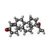  Mass: 330.461 Da / Num. of mol.: 2 / Source method: obtained synthetically / Formula: C21H30O3 Mass: 330.461 Da / Num. of mol.: 2 / Source method: obtained synthetically / Formula: C21H30O3#4: Chemical | ChemComp-NA / |   Mass: 22.990 Da / Num. of mol.: 1 / Source method: obtained synthetically / Formula: Na Mass: 22.990 Da / Num. of mol.: 1 / Source method: obtained synthetically / Formula: Na#5: Water | ChemComp-HOH / |  Mass: 18.015 Da / Num. of mol.: 239 / Source method: isolated from a natural source / Formula: H2O Mass: 18.015 Da / Num. of mol.: 239 / Source method: isolated from a natural source / Formula: H2O |
|---|
-Experimental details
-Experiment
| Experiment | Method: X-RAY DIFFRACTION / Number of used crystals: 1 |
|---|
- Sample preparation
| Crystal | Density Matthews: 2.87 Å3/Da / Density % sol: 57.17 % |
|---|---|
| Crystal grow | Temperature: 273 K / Method: vapor diffusion, hanging drop / pH: 7 Details: 2.5-2.8 M sodium acetate trihydrate, 0.1 M Bis-Tris propane, pH 7.0, VAPOR DIFFUSION, HANGING DROP, temperature 273K |
-Data collection
| Diffraction | Mean temperature: 100 K |
|---|---|
| Diffraction source | Source: SYNCHROTRON / Site: APS  / Beamline: 22-BM / Wavelength: 1 / Beamline: 22-BM / Wavelength: 1 |
| Detector | Type: MARMOSAIC 225 mm CCD / Detector: CCD / Date: Nov 8, 2008 |
| Radiation | Monochromator: SAGITALLY FOCUSED Si(111) / Protocol: SINGLE WAVELENGTH / Monochromatic (M) / Laue (L): M / Scattering type: x-ray |
| Radiation wavelength | Wavelength: 1 Å / Relative weight: 1 |
| Reflection | Resolution: 1.95→96.674 Å / Num. all: 48182 / Num. obs: 48012 / % possible obs: 93.9 % / Observed criterion σ(F): 0 / Observed criterion σ(I): 0 / Redundancy: 3.9 % / Biso Wilson estimate: 29.488 Å2 / Rsym value: 0.048 / Net I/σ(I): 13 |
| Reflection shell | Resolution: 1.95→2 Å / % possible all: 78.3 |
- Processing
| Software |
| ||||||||||||||||||||||||||||
|---|---|---|---|---|---|---|---|---|---|---|---|---|---|---|---|---|---|---|---|---|---|---|---|---|---|---|---|---|---|
| Refinement | Method to determine structure: MOLECULAR REPLACEMENT Starting model: PDB ENTRY 2Q3Y Resolution: 1.95→96.67 Å / Occupancy max: 1 / Occupancy min: 0.5 / σ(F): 0 / Stereochemistry target values: Engh & Huber
| ||||||||||||||||||||||||||||
| Displacement parameters | Biso max: 105.4 Å2 / Biso mean: 29.4878 Å2 / Biso min: 12.71 Å2 | ||||||||||||||||||||||||||||
| Refine analyze |
| ||||||||||||||||||||||||||||
| Refinement step | Cycle: LAST / Resolution: 1.95→96.67 Å
| ||||||||||||||||||||||||||||
| Refine LS restraints |
|
