 Movie
Movie Controller


+ Open data

- Basic information
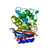
| Entry | Database: PDB / ID: 5hvi | |||||||||
|---|---|---|---|---|---|---|---|---|---|---|
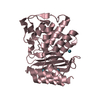
| Title | Crystal structure of TEM1 beta-lactamase | |||||||||
 Components Components | Beta-lactamase TEM | |||||||||
 Keywords Keywords | HYDROLASE | |||||||||
| Function / homology |  Function and homology information Function and homology informationAntimicrobial resistance / beta-lactam antibiotic catabolic process / beta-lactamase activity / beta-lactamase / response to antibiotic Similarity search - Function | |||||||||
| Biological species |  | |||||||||
| Method |  X-RAY DIFFRACTION / SYNCHROTRON / MOLECULAR REPLACEMENT / Resolution: 1.64 Å X-RAY DIFFRACTION / SYNCHROTRON / MOLECULAR REPLACEMENT / Resolution: 1.64 Å | |||||||||
 Authors Authors | Roose, B.W. / Dmochowski, I.J. | |||||||||
| Funding support |  United States, 2items United States, 2items
| |||||||||
 Citation Citation | Journal: Chemphyschem / Year: 2018 Title: A Structural Basis for129Xe Hyper-CEST Signal in TEM-1 beta-Lactamase. Authors: Roose, B.W. / Zemerov, S.D. / Wang, Y. / Kasimova, M.A. / Carnevale, V. / Dmochowski, I.J. | |||||||||
| History |
|
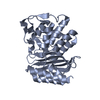
- Structure visualization
| Structure viewer | Molecule: MolmilJmol/JSmol |
|---|
- Downloads & links
-Download
| PDBx/mmCIF format | 5hvi.cif.gz | 413 KB | Display | PDBx/mmCIF format |
|---|---|---|---|---|
| PDB format | pdb5hvi.ent.gz | 335.5 KB | Display | PDB format |
| PDBx/mmJSON format | 5hvi.json.gz | Tree view | PDBx/mmJSON format | |
| Others |  Other downloads Other downloads |
-Validation report
| Arichive directory | https://data.pdbj.org/pub/pdb/validation_reports/hv/5hviftp://data.pdbj.org/pub/pdb/validation_reports/hv/5hvi | HTTPS FTP |
|---|
-Related structure data
| Related structure data |  5hw1C  5i52C  5i63C  5kkfC  5kpuC 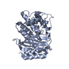 6apaC 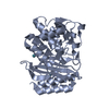 6aykC  1btlS C: citing same article ( S: Starting model for refinement |
|---|---|
| Similar structure data |


-Links
 PDBj
PDBj


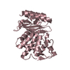
- Assembly
| Deposited unit | 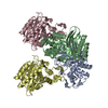
| ||||||||
|---|---|---|---|---|---|---|---|---|---|
| 1 | 
| ||||||||
| 2 | 
| ||||||||
| 3 | 
| ||||||||
| 4 | 
| ||||||||
| Unit cell |
|
-Components
| #1: Protein | Mass: 28911.904 Da / Num. of mol.: 4 / Mutation: M180T Source method: isolated from a genetically manipulated source Source: (gene. exp.) #2: Water | ChemComp-HOH / |  Mass: 18.015 Da / Num. of mol.: 967 / Source method: isolated from a natural source / Formula: H2O Mass: 18.015 Da / Num. of mol.: 967 / Source method: isolated from a natural source / Formula: H2OHas protein modification | Y | |
|---|
-Experimental details
-Experiment
| Experiment | Method: X-RAY DIFFRACTION / Number of used crystals: 1 |
|---|
- Sample preparation
| Crystal | Density Matthews: 2.11 Å3/Da / Density % sol: 41.77 % |
|---|---|
| Crystal grow | Temperature: 294 K / Method: vapor diffusion, hanging drop Details: 2% (v/v) tacsimate (pH 6.0), 0.1 M Bis-Tris (pH 6.5), 20% (w/v) PEG 3350 PH range: 6.0 - 7.0 |
-Data collection
| Diffraction | Mean temperature: 100 K |
|---|---|
| Diffraction source | Source: SYNCHROTRON / Site: ALS / Beamline: 4.2.2 / Wavelength: 1 Å |
| Detector | Type: NOIR-1 / Detector: CCD / Date: Dec 6, 2014 |
| Radiation | Protocol: SINGLE WAVELENGTH / Monochromatic (M) / Laue (L): M / Scattering type: x-ray |
| Radiation wavelength | Wavelength: 1 Å / Relative weight: 1 |
| Reflection twin | Operator: h,-k,-l / Fraction: 0.02 |
| Reflection | Resolution: 1.64→63.2 Å / Num. obs: 113836 / % possible obs: 96.9 % / Redundancy: 3.6 % / CC1/2: 0.998 / Rmerge(I) obs: 0.061 / Rpim(I) all: 0.037 / Net I/σ(I): 15.1 / Num. measured all: 409738 / Scaling rejects: 276 |
| Reflection shell | Resolution: 1.64→1.67 Å / Redundancy: 3.4 % / Rmerge(I) obs: 0.334 / Mean I/σ(I) obs: 3.4 / % possible all: 89.6 |
- Processing
| Software |
| |||||||||||||||||||||||||||||||||||||||||||||||||||||||||||||||||||||||||||||||||||||||||||||||||||||||||||||||||||||||||||||||||||||||||||||||||||
|---|---|---|---|---|---|---|---|---|---|---|---|---|---|---|---|---|---|---|---|---|---|---|---|---|---|---|---|---|---|---|---|---|---|---|---|---|---|---|---|---|---|---|---|---|---|---|---|---|---|---|---|---|---|---|---|---|---|---|---|---|---|---|---|---|---|---|---|---|---|---|---|---|---|---|---|---|---|---|---|---|---|---|---|---|---|---|---|---|---|---|---|---|---|---|---|---|---|---|---|---|---|---|---|---|---|---|---|---|---|---|---|---|---|---|---|---|---|---|---|---|---|---|---|---|---|---|---|---|---|---|---|---|---|---|---|---|---|---|---|---|---|---|---|---|---|---|---|---|
| Refinement | Method to determine structure: MOLECULAR REPLACEMENT Starting model: 1BTL Resolution: 1.64→63.198 Å / Cross valid method: FREE R-VALUE / σ(F): 1.96 / Phase error: 22.83
| |||||||||||||||||||||||||||||||||||||||||||||||||||||||||||||||||||||||||||||||||||||||||||||||||||||||||||||||||||||||||||||||||||||||||||||||||||
| Solvent computation | Shrinkage radii: 0.9 Å / VDW probe radii: 1.11 Å | |||||||||||||||||||||||||||||||||||||||||||||||||||||||||||||||||||||||||||||||||||||||||||||||||||||||||||||||||||||||||||||||||||||||||||||||||||
| Displacement parameters | Biso max: 50.22 Å2 / Biso mean: 12.2983 Å2 / Biso min: 3.87 Å2 | |||||||||||||||||||||||||||||||||||||||||||||||||||||||||||||||||||||||||||||||||||||||||||||||||||||||||||||||||||||||||||||||||||||||||||||||||||
| Refinement step | Cycle: final / Resolution: 1.64→63.198 Å
| |||||||||||||||||||||||||||||||||||||||||||||||||||||||||||||||||||||||||||||||||||||||||||||||||||||||||||||||||||||||||||||||||||||||||||||||||||
| Refine LS restraints |
| |||||||||||||||||||||||||||||||||||||||||||||||||||||||||||||||||||||||||||||||||||||||||||||||||||||||||||||||||||||||||||||||||||||||||||||||||||
| LS refinement shell | Refine-ID: X-RAY DIFFRACTION / Total num. of bins used: 20
|
