 Movie
Movie Controller


+ Open data

- Basic information
| Entry | Database: PDB / ID: 1htz | ||||||
|---|---|---|---|---|---|---|---|
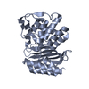
| Title | CRYSTAL STRUCTURE OF TEM52 BETA-LACTAMASE | ||||||
 Components Components | BETA-LACTAMASE MUTANT TEM52 | ||||||
 Keywords Keywords | HYDROLASE / Mutant form of beta-lactamase | ||||||
| Function / homology |  Function and homology information Function and homology informationbeta-lactam antibiotic catabolic process / beta-lactamase activity / beta-lactamase / response to antibiotic Similarity search - Function | ||||||
| Biological species |  Klebsiella pneumoniae (bacteria) Klebsiella pneumoniae (bacteria) | ||||||
| Method |  X-RAY DIFFRACTION / SYNCHROTRON / MOLECULAR REPLACEMENT / Resolution: 2.4 Å X-RAY DIFFRACTION / SYNCHROTRON / MOLECULAR REPLACEMENT / Resolution: 2.4 Å | ||||||
 Authors Authors | Stevens, R.C. / Orencia, M.C. | ||||||
 Citation Citation | Journal: Nat.Struct.Biol. / Year: 2001 Title: Predicting the emergence of antibiotic resistance by directed evolution and structural analysis. Authors: Orencia, M.C. / Yoon, J.S. / Ness, J.E. / Stemmer, W.P. / Stevens, R.C. | ||||||
| History |
|
- Structure visualization
| Structure viewer | Molecule: MolmilJmol/JSmol |
|---|
- Downloads & links
-Download
| PDBx/mmCIF format | 1htz.cif.gz | 306.2 KB | Display | PDBx/mmCIF format |
|---|---|---|---|---|
| PDB format | pdb1htz.ent.gz | 252.4 KB | Display | PDB format |
| PDBx/mmJSON format | 1htz.json.gz | Tree view | PDBx/mmJSON format | |
| Others |  Other downloads Other downloads |
-Validation report
| Arichive directory | https://data.pdbj.org/pub/pdb/validation_reports/ht/1htzftp://data.pdbj.org/pub/pdb/validation_reports/ht/1htz | HTTPS FTP |
|---|
-Related structure data
| Related structure data |  1temS S: Starting model for refinement |
|---|---|
| Similar structure data |


-Links
 PDBj
PDBj


- Assembly
| Deposited unit | 
| ||||||||
|---|---|---|---|---|---|---|---|---|---|
| 1 | 
| ||||||||
| 2 | 
| ||||||||
| 3 | 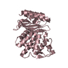
| ||||||||
| 4 | 
| ||||||||
| 5 | 
| ||||||||
| 6 | 
| ||||||||
| Unit cell |
|
-Components
| #1: Protein | Mass: 28941.998 Da / Num. of mol.: 6 Source method: isolated from a genetically manipulated source Source: (gene. exp.) Klebsiella pneumoniae (bacteria) / Plasmid: PET30A / Species (production host): Escherichia coli / Production host: #2: Water | ChemComp-HOH / |  Mass: 18.015 Da / Num. of mol.: 585 / Source method: isolated from a natural source / Formula: H2O Mass: 18.015 Da / Num. of mol.: 585 / Source method: isolated from a natural source / Formula: H2OHas protein modification | Y | |
|---|
-Experimental details
-Experiment
| Experiment | Method: X-RAY DIFFRACTION / Number of used crystals: 3 |
|---|
- Sample preparation
| Crystal | Density Matthews: 2.81 Å3/Da / Density % sol: 56.27 % | ||||||||||||||||||||||||||||||
|---|---|---|---|---|---|---|---|---|---|---|---|---|---|---|---|---|---|---|---|---|---|---|---|---|---|---|---|---|---|---|---|
| Crystal grow | Temperature: 293 K / Method: vapor diffusion, hanging drop / pH: 6.5 Details: 16% PEG8K, 125 mM MgOAc, 25mM Na citrate, pH 6.5, VAPOR DIFFUSION, HANGING DROP, temperature 20K | ||||||||||||||||||||||||||||||
| Crystal grow | *PLUS | ||||||||||||||||||||||||||||||
| Components of the solutions | *PLUS
|
-Data collection
| Diffraction | Mean temperature: 100 K |
|---|---|
| Diffraction source | Source: SYNCHROTRON / Site: ALS  / Beamline: 5.0.3 / Wavelength: 1 / Beamline: 5.0.3 / Wavelength: 1 |
| Detector | Type: ADSC QUANTUM 4 / Detector: CCD / Date: Jan 15, 1999 / Details: mirrors |
| Radiation | Monochromator: ALS 5.0.2 / Protocol: SINGLE WAVELENGTH / Monochromatic (M) / Laue (L): M / Scattering type: x-ray |
| Radiation wavelength | Wavelength: 1 Å / Relative weight: 1 |
| Reflection | Resolution: 2.4→20 Å / Num. all: 73211 / Num. obs: 69960 / % possible obs: 91 % / Observed criterion σ(F): 2 / Observed criterion σ(I): 2 / Redundancy: 10 % / Biso Wilson estimate: 43.5 Å2 / Rmerge(I) obs: 0.115 / Net I/σ(I): 22.4 |
| Reflection shell | Resolution: 2.4→2.5 Å / Redundancy: 6 % / Rmerge(I) obs: 0.432 / Mean I/σ(I) obs: 7.1 / % possible all: 80 |
| Reflection | *PLUS Num. measured all: 736906 |
| Reflection shell | *PLUS % possible obs: 80 % |
- Processing
| Software |
| |||||||||||||||||||||||||
|---|---|---|---|---|---|---|---|---|---|---|---|---|---|---|---|---|---|---|---|---|---|---|---|---|---|---|
| Refinement | Method to determine structure: MOLECULAR REPLACEMENT Starting model: 1TEM Resolution: 2.4→20 Å / Isotropic thermal model: isotropic / Cross valid method: THROUGHOUT / σ(F): 2 / σ(I): 2 / Stereochemistry target values: Engh & Huber Details: 5 of 6 molecules refined using NCS, 6th molecule different due to packing. Pseudo-6 fold symmetry, attempts to process data as hexagonal or rhombohedral were unsuccessful
| |||||||||||||||||||||||||
| Displacement parameters | Biso mean: 51 Å2 | |||||||||||||||||||||||||
| Refinement step | Cycle: LAST / Resolution: 2.4→20 Å
| |||||||||||||||||||||||||
| Refine LS restraints |
| |||||||||||||||||||||||||
| Software | *PLUS Name: CNS / Version: 0.9 / Classification: refinement | |||||||||||||||||||||||||
| Refine LS restraints | *PLUS Type: c_bond_d / Dev ideal: 0.006 |
