 Movie
Movie Controller


+ Open data

- Basic information
| Entry | Database: PDB / ID: 4gku | ||||||
|---|---|---|---|---|---|---|---|
| Title | Crystal structure of beta lactamase in PET-15B | ||||||
 Components Components | Beta-lactamase TEM | ||||||
 Keywords Keywords | HYDROLASE / beta lactamase | ||||||
| Function / homology |  Function and homology information Function and homology informationAntimicrobial resistance / beta-lactam antibiotic catabolic process / beta-lactamase activity / beta-lactamase / response to antibiotic Similarity search - Function | ||||||
| Biological species |  | ||||||
| Method |  X-RAY DIFFRACTION / SYNCHROTRON / MOLECULAR REPLACEMENT / Resolution: 1.915 Å X-RAY DIFFRACTION / SYNCHROTRON / MOLECULAR REPLACEMENT / Resolution: 1.915 Å | ||||||
 Authors Authors | Zhang, Y. / Cao, C. | ||||||
 Citation Citation | Journal: To be published Title: Crystal structure of beta lactamase in PET-15B Authors: Zhang, Y. / Cao, C. | ||||||
| History |
|
- Structure visualization
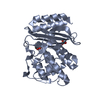
| Structure viewer | Molecule: MolmilJmol/JSmol |
|---|
- Downloads & links
-Download
| PDBx/mmCIF format | 4gku.cif.gz | 70.1 KB | Display | PDBx/mmCIF format |
|---|---|---|---|---|
| PDB format | pdb4gku.ent.gz | 50.5 KB | Display | PDB format |
| PDBx/mmJSON format | 4gku.json.gz | Tree view | PDBx/mmJSON format | |
| Others |  Other downloads Other downloads |
-Validation report
| Arichive directory | https://data.pdbj.org/pub/pdb/validation_reports/gk/4gkuftp://data.pdbj.org/pub/pdb/validation_reports/gk/4gku | HTTPS FTP |
|---|
-Related structure data
| Similar structure data |
|---|


-Links
 PDBj
PDBj


- Assembly
| Deposited unit | 
| ||||||||
|---|---|---|---|---|---|---|---|---|---|
| 1 |
| ||||||||
| Unit cell |
|
-Components
| #1: Protein | Mass: 28941.994 Da / Num. of mol.: 1 Source method: isolated from a genetically manipulated source Source: (gene. exp.) |
|---|---|
| #2: Water | ChemComp-HOH /  Mass: 18.015 Da / Num. of mol.: 315 / Source method: isolated from a natural source / Formula: H2O Mass: 18.015 Da / Num. of mol.: 315 / Source method: isolated from a natural source / Formula: H2O |
| Has protein modification | Y |
-Experimental details
-Experiment
| Experiment | Method: X-RAY DIFFRACTION / Number of used crystals: 1 |
|---|
- Sample preparation
| Crystal | Density Matthews: 2.21 Å3/Da / Density % sol: 44.33 % |
|---|---|
| Crystal grow | Temperature: 293 K / Method: vapor diffusion / pH: 6.5 Details: 0.2M magnesium chloride, 0.1M BIS-Tris, 25% PEG 3350, pH 6.5, VAPOR DIFFUSION, temperature 293K |
-Data collection
| Diffraction | Mean temperature: 100 K |
|---|---|
| Diffraction source | Source: SYNCHROTRON / Site: BSRF  / Beamline: 3W1A / Wavelength: 1.2605 Å / Beamline: 3W1A / Wavelength: 1.2605 Å |
| Detector | Type: MAR CCD 165 mm / Detector: CCD / Date: Jul 25, 2010 |
| Radiation | Protocol: SINGLE WAVELENGTH / Monochromatic (M) / Laue (L): M / Scattering type: x-ray |
| Radiation wavelength | Wavelength: 1.2605 Å / Relative weight: 1 |
| Reflection | Resolution: 1.91→50 Å / Num. all: 37743 / Num. obs: 37743 / % possible obs: 98.1 % / Observed criterion σ(F): 2.3 / Observed criterion σ(I): 100.8 / Redundancy: 13.1 % |
| Reflection shell | Resolution: 1.91→1.98 Å / % possible all: 89.3 |
- Processing
| Software |
| ||||||||||||||||||||||||||||||||||||||||||||||||||||||||||||||||||
|---|---|---|---|---|---|---|---|---|---|---|---|---|---|---|---|---|---|---|---|---|---|---|---|---|---|---|---|---|---|---|---|---|---|---|---|---|---|---|---|---|---|---|---|---|---|---|---|---|---|---|---|---|---|---|---|---|---|---|---|---|---|---|---|---|---|---|---|
| Refinement | Method to determine structure: MOLECULAR REPLACEMENT / Resolution: 1.915→34.917 Å / SU ML: 0.19 / σ(F): 0 / Phase error: 17.83 / Stereochemistry target values: MLHL
| ||||||||||||||||||||||||||||||||||||||||||||||||||||||||||||||||||
| Solvent computation | Shrinkage radii: 0.95 Å / VDW probe radii: 1.2 Å / Solvent model: FLAT BULK SOLVENT MODEL / Bsol: 40 Å2 / ksol: 0.4 e/Å3 | ||||||||||||||||||||||||||||||||||||||||||||||||||||||||||||||||||
| Displacement parameters |
| ||||||||||||||||||||||||||||||||||||||||||||||||||||||||||||||||||
| Refinement step | Cycle: LAST / Resolution: 1.915→34.917 Å
| ||||||||||||||||||||||||||||||||||||||||||||||||||||||||||||||||||
| Refine LS restraints |
| ||||||||||||||||||||||||||||||||||||||||||||||||||||||||||||||||||
| LS refinement shell | Refine-ID: X-RAY DIFFRACTION / Total num. of bins used: 10
|
