 Movie
Movie Controller

[English] 日本語
 Yorodumi
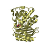
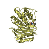
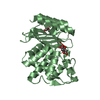
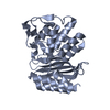
- PDB-1tem: 6 ALPHA HYDROXYMETHYL PENICILLOIC ACID ACYLATED ON THE TEM-1 BETA... -
+ Open data

- Basic information
| Entry | Database: PDB / ID: 1tem | ||||||
|---|---|---|---|---|---|---|---|
| Title | 6 ALPHA HYDROXYMETHYL PENICILLOIC ACID ACYLATED ON THE TEM-1 BETA-LACTAMASE FROM ESCHERICHIA COLI | ||||||
 Components Components | TEM-1 BETA LACTAMASE | ||||||
 Keywords Keywords | HYDROLASE / ANTIBIOTIC RESISTANCE / TRANSPOSABLE ELEMENT | ||||||
| Function / homology |  Function and homology information Function and homology informationAntimicrobial resistance / beta-lactam antibiotic catabolic process / beta-lactamase activity / beta-lactamase / response to antibiotic Similarity search - Function | ||||||
| Biological species |  | ||||||
| Method |  X-RAY DIFFRACTION / SYNCHROTRON / Resolution: 1.95 Å X-RAY DIFFRACTION / SYNCHROTRON / Resolution: 1.95 Å | ||||||
 Authors Authors | Maveyraud, L. / Massova, I. / Samama, J.P. / Mobashery, S. | ||||||
 Citation Citation | Journal: J.Am.Chem.Soc. / Year: 1996 Title: Crystal Structure of 6Alpha-Hydroxymethylpenicillanate Complexed to the Tem-1 Beta-Lactamase from Escherichia Coli: Evidence on the Mechanism of Action of a Novel Inhibitor Designed by a Computer-Aided Process Authors: Maveyraud, L. / Massova, I. / Birck, C. / Miyashita, K. / Samama, J.P. / Mobashery, S. #1: Journal: J.Am.Chem.Soc. / Year: 1995Title: Design, Synthesis, and Evaluation of a Potent Mechanism-Based Inhibitor for the Tem Beta-Lactamase with Implications for the Enzyme Mechanism Authors: Miyashita, K. / Massova, I. / Taibi, P. / Mobashery, S. | ||||||
| History |
|
- Structure visualization
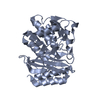
| Structure viewer | Molecule: MolmilJmol/JSmol |
|---|
- Downloads & links
-Download
| PDBx/mmCIF format | 1tem.cif.gz | 69.9 KB | Display | PDBx/mmCIF format |
|---|---|---|---|---|
| PDB format | pdb1tem.ent.gz | 49.9 KB | Display | PDB format |
| PDBx/mmJSON format | 1tem.json.gz | Tree view | PDBx/mmJSON format | |
| Others |  Other downloads Other downloads |
-Validation report
| Arichive directory | https://data.pdbj.org/pub/pdb/validation_reports/te/1temftp://data.pdbj.org/pub/pdb/validation_reports/te/1tem | HTTPS FTP |
|---|
-Related structure data
| Similar structure data |
|---|


-Links
 PDBj
PDBj


- Assembly
| Deposited unit | 
| ||||||||
|---|---|---|---|---|---|---|---|---|---|
| 1 |
| ||||||||
| Unit cell |
|
-Components
| #1: Protein | Mass: 28984.076 Da / Num. of mol.: 1 Source method: isolated from a genetically manipulated source Details: VARIANT V84I, A184V,ACYL-ENZYME COMPLEX / Source: (gene. exp.) |
|---|---|
| #2: Chemical | ChemComp-ALP / 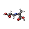  Mass: 249.284 Da / Num. of mol.: 1 / Source method: obtained synthetically / Formula: C9H15NO5S Mass: 249.284 Da / Num. of mol.: 1 / Source method: obtained synthetically / Formula: C9H15NO5S |
| #3: Water | ChemComp-HOH /  Mass: 18.015 Da / Num. of mol.: 240 / Source method: isolated from a natural source / Formula: H2O Mass: 18.015 Da / Num. of mol.: 240 / Source method: isolated from a natural source / Formula: H2O |
| Compound details | THE STRUCTURE DEPOSITED IS AN ACYL-ENZYME COMPLEX, BETWEEN 6ALPHA-HYDROXYMETHYL PENICILLOIC ACID ...THE STRUCTURE DEPOSITED IS AN ACYL-ENZYME COMPLEX, BETWEEN 6ALPHA-HYDROXYMET |
| Has protein modification | Y |
-Experimental details
-Experiment
| Experiment | Method: X-RAY DIFFRACTION / Number of used crystals: 1 |
|---|
- Sample preparation
| Crystal | Density Matthews: 2.03 Å3/Da / Density % sol: 39.52 % | |||||||||||||||||||||||||||||||||||
|---|---|---|---|---|---|---|---|---|---|---|---|---|---|---|---|---|---|---|---|---|---|---|---|---|---|---|---|---|---|---|---|---|---|---|---|---|
| Crystal grow | pH: 7.8 / Details: pH 7.8 | |||||||||||||||||||||||||||||||||||
| Crystal grow | *PLUS Temperature: 6 ℃ / Method: vapor diffusion, hanging drop / Details: Jelsch, C., (1992) J. Mol. Biol., 223, 377. | |||||||||||||||||||||||||||||||||||
| Components of the solutions | *PLUS
|
-Data collection
| Diffraction | Mean temperature: 263 K |
|---|---|
| Diffraction source | Source: SYNCHROTRON / Site: EMBL/DESY, HAMBURG  / Beamline: X31 / Wavelength: 0.933 / Beamline: X31 / Wavelength: 0.933 |
| Detector | Type: MARRESEARCH / Detector: IMAGE PLATE / Date: Dec 15, 1995 |
| Radiation | Monochromatic (M) / Laue (L): M / Scattering type: x-ray |
| Radiation wavelength | Wavelength: 0.933 Å / Relative weight: 1 |
| Reflection | Num. obs: 17104 / % possible obs: 96.2 % / Observed criterion σ(I): 0 / Redundancy: 2.5 % / Biso Wilson estimate: 12.49 Å2 / Rmerge(I) obs: 0.046 |
| Reflection | *PLUS Highest resolution: 1.95 Å / Lowest resolution: 22 Å / Num. measured all: 42284 |
| Reflection shell | *PLUS Highest resolution: 1.95 Å / Lowest resolution: 2.02 Å / % possible obs: 94.8 % / Num. unique obs: 1524 / Num. measured obs: 3622 / Rmerge(I) obs: 0.076 |
- Processing
| Software |
| ||||||||||||||||||||||||||||||||||||||||||||||||||||||||||||
|---|---|---|---|---|---|---|---|---|---|---|---|---|---|---|---|---|---|---|---|---|---|---|---|---|---|---|---|---|---|---|---|---|---|---|---|---|---|---|---|---|---|---|---|---|---|---|---|---|---|---|---|---|---|---|---|---|---|---|---|---|---|
| Refinement | Resolution: 1.95→8 Å / σ(F): 0
| ||||||||||||||||||||||||||||||||||||||||||||||||||||||||||||
| Displacement parameters | Biso mean: 12.35 Å2 | ||||||||||||||||||||||||||||||||||||||||||||||||||||||||||||
| Refine analyze | Luzzati d res low obs: 8 Å / Luzzati sigma a obs: 0.11 Å | ||||||||||||||||||||||||||||||||||||||||||||||||||||||||||||
| Refinement step | Cycle: LAST / Resolution: 1.95→8 Å
| ||||||||||||||||||||||||||||||||||||||||||||||||||||||||||||
| Refine LS restraints |
| ||||||||||||||||||||||||||||||||||||||||||||||||||||||||||||
| LS refinement shell | Resolution: 1.95→1.98 Å
| ||||||||||||||||||||||||||||||||||||||||||||||||||||||||||||
| Software | *PLUS Name: X-PLOR / Version: 3.1 / Classification: refinement | ||||||||||||||||||||||||||||||||||||||||||||||||||||||||||||
| Refinement | *PLUS | ||||||||||||||||||||||||||||||||||||||||||||||||||||||||||||
| Solvent computation | *PLUS | ||||||||||||||||||||||||||||||||||||||||||||||||||||||||||||
| Displacement parameters | *PLUS Biso mean: 12.359 Å2 |
