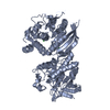
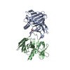
- PDB-2y7i: Structural basis for high arginine specificity in Salmonella typh... -
+
Open data
ID or keywords:
Loading...
-
Basic information
Entry
Database: PDB / ID: 2y7i
Title
Structural basis for high arginine specificity in Salmonella typhimurium periplasmic binding protein STM4351.
Components
STM4351
Keywords
ARGININE-BINDING PROTEIN
Function / homology
Function and homology information
amino acid binding / ligand-gated monoatomic ion channel activity / outer membrane-bounded periplasmic space / membrane / metal ion binding Similarity search - Function
Solute-binding protein family 3, conserved site / Bacterial extracellular solute-binding proteins, family 3 signature. / Bacterial extracellular solute-binding proteins, family 3 / Solute-binding protein family 3/N-terminal domain of MltF / Bacterial periplasmic substrate-binding proteins / Ionotropic glutamate receptor / Eukaryotic homologues of bacterial periplasmic substrate binding proteins. / Periplasmic binding protein-like II / D-Maltodextrin-Binding Protein; domain 2 / 3-Layer(aba) Sandwich / Alpha Beta Similarity search - Domain/homology
Resolution: 1.9→47.28 Å / Cor.coef. Fo:Fc: 0.951 / Cor.coef. Fo:Fc free: 0.936 / SU B: 7.634 / SU ML: 0.109 / Cross valid method: THROUGHOUT / ESU R: 0.17 / ESU R Free: 0.145 / Stereochemistry target values: MAXIMUM LIKELIHOOD / Details: HYDROGENS HAVE BEEN ADDED IN THE RIDING POSITIONS.
Rfactor
Num. reflection
% reflection
Selection details
Rfree
0.22602
1856
5 %
RANDOM
Rwork
0.19805
-
-
-
obs
0.19949
35156
99.87 %
-
Solvent computation
Ion probe radii: 0.8 Å / Shrinkage radii: 0.8 Å / VDW probe radii: 1.4 Å / Solvent model: MASK
 Movie
Movie Controller

 Yorodumi
 Open data
 Basic information
 Components
 Keywords
 Function and homology information
 SALMONELLA ENTERICA SUBSP. ENTERICA (bacteria)
 X-RAY DIFFRACTION /
  Authors
 Citation
 Structure visualization
 Downloads & links
 Other downloads
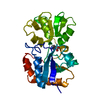


 PDBj
PDBj

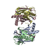
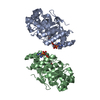
 Assembly
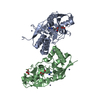


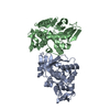
 Type: L-peptide linking / Mass: 175.209 Da / Num. of mol.: 2 / Source method: obtained synthetically / Formula: C6H15N4O2
 Mass: 65.409 Da / Num. of mol.: 6 / Source method: obtained synthetically / Formula: Zn
 Mass: 59.044 Da / Num. of mol.: 2 / Source method: obtained synthetically / Formula: C2H3O2
 Mass: 92.094 Da / Num. of mol.: 2 / Source method: obtained synthetically / Formula: C3H8O3
 Sample preparation
 Processing