 Movie
Movie Controller

[English] 日本語
 Yorodumi
- PDB-1jif: Crystal structure of bleomycin-binding protein from bleomycin-pro... -
+ Open data

- Basic information
| Entry | Database: PDB / ID: 1jif | ||||||
|---|---|---|---|---|---|---|---|
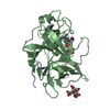
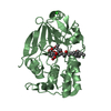
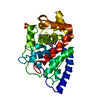
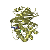
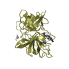
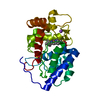
| Title | Crystal structure of bleomycin-binding protein from bleomycin-producing Streptomyces verticillus complexed with copper(II)-bleomycin | ||||||
 Components Components | bleomycin-binding protein | ||||||
 Keywords Keywords | PROTEIN BINDING / protein-ligand complex | ||||||
| Function / homology |  Function and homology information Function and homology information | ||||||
| Biological species |  Streptomyces verticillus (bacteria) Streptomyces verticillus (bacteria) | ||||||
| Method |  X-RAY DIFFRACTION / MOLECULAR REPLACEMENT / Resolution: 1.6 Å X-RAY DIFFRACTION / MOLECULAR REPLACEMENT / Resolution: 1.6 Å | ||||||
 Authors Authors | Sugiyama, M. / Kumagai, T. / Hayashida, M. / Maruyama, M. / Matoba, Y. | ||||||
 Citation Citation | Journal: J.Biol.Chem. / Year: 2002 Title: The 1.6-A crystal structure of the copper(II)-bound bleomycin complexed with the bleomycin-binding protein from bleomycin-producing Streptomyces verticillus. Authors: Sugiyama, M. / Kumagai, T. / Hayashida, M. / Maruyama, M. / Matoba, Y. #1: Journal: J.Mol.Biol. / Year: 2000Title: The 1.5 A crystal structure of a bleomycin resistance determinant from bleomycin-producing Streptomyces verticillus Authors: Kawano, Y. / Kumagai, T. / Muta, K. / Matoba, Y. / Davies, J. / Sugiyama, M. #2: Journal: J.Biol.Chem. / Year: 2001Title: Crystal structures of the transposon Tn5-carried bleomycin resistance determinant uncomplexed and complexed with bleomycin Authors: Maruyama, M. / Kumagai, T. / Matoba, Y. / Hayashida, M. / Fujii, T. / Hata, Y. / Sugiyama, M. #3: Journal: Acta Crystallogr.,Sect.D / Year: 1998Title: Crystallization and preliminary X-ray diffraction studies of bleomycin-binding protein from bleomycin-producing Streptomyces verticillus Authors: Kumagai, T. / Muta, K. / Matoba, Y. / Kawano, Y. / Kamiya, N. / Davies, J. / Sugiyama, M. | ||||||
| History |
|
- Structure visualization
| Structure viewer | Molecule: MolmilJmol/JSmol |
|---|
- Downloads & links
-Download
| PDBx/mmCIF format | 1jif.cif.gz | 70.9 KB | Display | PDBx/mmCIF format |
|---|---|---|---|---|
| PDB format | pdb1jif.ent.gz | 51.7 KB | Display | PDB format |
| PDBx/mmJSON format | 1jif.json.gz | Tree view | PDBx/mmJSON format | |
| Others |  Other downloads Other downloads |
-Validation report
| Arichive directory | https://data.pdbj.org/pub/pdb/validation_reports/ji/1jifftp://data.pdbj.org/pub/pdb/validation_reports/ji/1jif | HTTPS FTP |
|---|
-Related structure data


-Links
 PDBj
PDBj- Assembly
| Deposited unit | 
| ||||||||
|---|---|---|---|---|---|---|---|---|---|
| 1 |
| ||||||||
| Unit cell |
| ||||||||
| Details | The biological assembly is a homodimer. |
-Components
| #1: Protein | Mass: 13243.607 Da / Num. of mol.: 2 Source method: isolated from a genetically manipulated source Source: (gene. exp.) Streptomyces verticillus (bacteria) / Gene: blmA / Plasmid: pKKtrp / Production host: #2: Chemical |   Mass: 63.546 Da / Num. of mol.: 2 / Source method: obtained synthetically / Formula: Cu Mass: 63.546 Da / Num. of mol.: 2 / Source method: obtained synthetically / Formula: Cu#3: Chemical |   Mass: 35.453 Da / Num. of mol.: 2 / Source method: obtained synthetically / Formula: Cl Mass: 35.453 Da / Num. of mol.: 2 / Source method: obtained synthetically / Formula: Cl#4: Chemical | 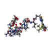  Mass: 1416.560 Da / Num. of mol.: 2 / Source method: obtained synthetically / Formula: C55H85N17O21S3 / Comment: medication*YM Mass: 1416.560 Da / Num. of mol.: 2 / Source method: obtained synthetically / Formula: C55H85N17O21S3 / Comment: medication*YM#5: Water | ChemComp-HOH / |  Mass: 18.015 Da / Num. of mol.: 117 / Source method: isolated from a natural source / Formula: H2O Mass: 18.015 Da / Num. of mol.: 117 / Source method: isolated from a natural source / Formula: H2O |
|---|
-Experimental details
-Experiment
| Experiment | Method: X-RAY DIFFRACTION / Number of used crystals: 1 |
|---|
- Sample preparation
| Crystal | Density Matthews: 1.92 Å3/Da / Density % sol: 36 % | ||||||||||||||||||||||||
|---|---|---|---|---|---|---|---|---|---|---|---|---|---|---|---|---|---|---|---|---|---|---|---|---|---|
| Crystal grow | Temperature: 298 K / Method: vapor diffusion, hanging drop / pH: 6 Details: PEG4000, ammonium acetate, sodium cacodylate, pH 6.0, VAPOR DIFFUSION, HANGING DROP, temperature 298K | ||||||||||||||||||||||||
| Crystal grow | *PLUS Temperature: 25 ℃ | ||||||||||||||||||||||||
| Components of the solutions | *PLUS
|
-Data collection
| Diffraction | Mean temperature: 293 K |
|---|---|
| Diffraction source | Source: ROTATING ANODE / Type: RIGAKU RU300 / Wavelength: 1.5418 Å |
| Detector | Type: RIGAKU RAXIS IIC / Detector: IMAGE PLATE / Date: Apr 1, 2000 |
| Radiation | Monochromator: graphite / Protocol: SINGLE WAVELENGTH / Monochromatic (M) / Laue (L): M / Scattering type: x-ray |
| Radiation wavelength | Wavelength: 1.5418 Å / Relative weight: 1 |
| Reflection | Resolution: 1.6→100 Å / Num. obs: 30741 / % possible obs: 96.7 % / Observed criterion σ(F): 2 / Observed criterion σ(I): 1 / Redundancy: 4.29 % / Rmerge(I) obs: 0.059 / Net I/σ(I): 18.4 |
| Reflection shell | Resolution: 1.6→1.7 Å / Rmerge(I) obs: 0.242 / Mean I/σ(I) obs: 1.98 / % possible all: 84.5 |
| Reflection | *PLUS Lowest resolution: 100 Å |
- Processing
| Software |
| |||||||||||||||||||||||||||||||||
|---|---|---|---|---|---|---|---|---|---|---|---|---|---|---|---|---|---|---|---|---|---|---|---|---|---|---|---|---|---|---|---|---|---|---|
| Refinement | Method to determine structure: MOLECULAR REPLACEMENT / Resolution: 1.6→10 Å / Num. parameters: 9018 / Num. restraintsaints: 8881 / Cross valid method: FREE R / σ(F): 2 / σ(I): 1 / Stereochemistry target values: Engh & Huber
| |||||||||||||||||||||||||||||||||
| Refine analyze | Num. disordered residues: 13 / Occupancy sum hydrogen: 0 / Occupancy sum non hydrogen: 2180 | |||||||||||||||||||||||||||||||||
| Refinement step | Cycle: LAST / Resolution: 1.6→10 Å
| |||||||||||||||||||||||||||||||||
| Refine LS restraints |
| |||||||||||||||||||||||||||||||||
| Software | *PLUS Name: SHELXL-97 / Classification: refinement | |||||||||||||||||||||||||||||||||
| Refinement | *PLUS Highest resolution: 1.6 Å / Lowest resolution: 10 Å / σ(F): 2 / % reflection Rfree: 10 % / Rfactor all: 0.15 / Rfactor obs: 0.159 / Rfactor Rfree: 0.208 / Rfactor Rwork: 0.158 | |||||||||||||||||||||||||||||||||
| Solvent computation | *PLUS | |||||||||||||||||||||||||||||||||
| Displacement parameters | *PLUS | |||||||||||||||||||||||||||||||||
| Refine LS restraints | *PLUS
|
