[English] 日本語
Yorodumi
- PDB-2vnx: Crystal structure of soybean ascorbate peroxidase mutant W41A aft... -

+
Open data


ID or keywords:

Loading...

-
Basic information

Entry
Database: PDB / ID: 2vnx
TitleCrystal structure of soybean ascorbate peroxidase mutant W41A after exposure to a high dose of x-rays
ComponentsASCORBATE PEROXIDASE
KeywordsOXIDOREDUCTASE / APX / PEROXIDASE / HEME ENZYME / REDUCTION BY X-RAYS
Function / homology
Function and homology information


L-ascorbate peroxidase / L-ascorbate peroxidase activity / cellular response to oxidative stress / heme binding / metal ion binding
Similarity search - Function
Class I peroxidase / Heme-binding peroxidase Ccp1-like / Peroxidase; domain 2 / Peroxidase, domain 2 / Peroxidase; domain 1 - #10 / Peroxidase; domain 1 / Peroxidases heam-ligand binding site / Peroxidase, active site / Peroxidases active site signature. / Plant heme peroxidase family profile. ...Class I peroxidase / Heme-binding peroxidase Ccp1-like / Peroxidase; domain 2 / Peroxidase, domain 2 / Peroxidase; domain 1 - #10 / Peroxidase; domain 1 / Peroxidases heam-ligand binding site / Peroxidase, active site / Peroxidases active site signature. / Plant heme peroxidase family profile. / Haem peroxidase / Peroxidase / Peroxidases proximal heme-ligand signature. / Haem peroxidase superfamily / Orthogonal Bundle / Mainly Alpha
Similarity search - Domain/homology
PROTOPORPHYRIN IX CONTAINING FE / L-ascorbate peroxidase
Similarity search - Component
Biological speciesGLYCINE MAX (soybean)
MethodX-RAY DIFFRACTION / SYNCHROTRON / MOLECULAR REPLACEMENT / Resolution: 1.5 Å
AuthorsMetcalfe, C.L. / Badyal, S.K. / Raven, E.L. / Moody, P.C.E.
CitationJournal: Biochemistry / Year: 2008
Title: Iron Oxidation State Modulates Active Site Structure in a Heme Peroxidase.
Authors: Badyal, S.K. / Metcalfe, C.L. / Basran, J. / Efimov, I. / Moody, P.C.E. / Raven, E.L.
History
DepositionFeb 8, 2008Deposition site: PDBE / Processing site: PDBE
Revision 1.0Apr 8, 2008Provider: repository / Type: Initial release
Revision 1.1May 8, 2011Group: Version format compliance
Revision 1.2Jul 13, 2011Group: Version format compliance
Revision 1.3Apr 10, 2019Group: Data collection / Other / Source and taxonomy
Category: entity_src_gen / pdbx_database_proc / pdbx_database_status
Item: _entity_src_gen.pdbx_host_org_cell_line / _entity_src_gen.pdbx_host_org_variant / _pdbx_database_status.recvd_author_approval
Revision 1.4Dec 13, 2023Group: Data collection / Database references ...Data collection / Database references / Derived calculations / Other / Refinement description
Category: chem_comp_atom / chem_comp_bond ...chem_comp_atom / chem_comp_bond / database_2 / pdbx_database_status / pdbx_initial_refinement_model / pdbx_struct_conn_angle / struct_conn / struct_site
Item: _database_2.pdbx_DOI / _database_2.pdbx_database_accession ..._database_2.pdbx_DOI / _database_2.pdbx_database_accession / _pdbx_database_status.status_code_sf / _pdbx_struct_conn_angle.ptnr1_auth_seq_id / _pdbx_struct_conn_angle.ptnr1_label_alt_id / _pdbx_struct_conn_angle.ptnr1_label_seq_id / _pdbx_struct_conn_angle.ptnr3_auth_seq_id / _pdbx_struct_conn_angle.ptnr3_label_alt_id / _pdbx_struct_conn_angle.ptnr3_label_seq_id / _pdbx_struct_conn_angle.value / _struct_conn.pdbx_dist_value / _struct_conn.pdbx_ptnr1_label_alt_id / _struct_conn.pdbx_ptnr2_label_alt_id / _struct_conn.ptnr1_auth_comp_id / _struct_conn.ptnr1_auth_seq_id / _struct_conn.ptnr1_label_asym_id / _struct_conn.ptnr1_label_atom_id / _struct_conn.ptnr1_label_comp_id / _struct_conn.ptnr1_label_seq_id / _struct_conn.ptnr2_auth_comp_id / _struct_conn.ptnr2_auth_seq_id / _struct_conn.ptnr2_label_asym_id / _struct_conn.ptnr2_label_atom_id / _struct_conn.ptnr2_label_comp_id / _struct_conn.ptnr2_label_seq_id / _struct_site.pdbx_auth_asym_id / _struct_site.pdbx_auth_comp_id / _struct_site.pdbx_auth_seq_id

-
Structure visualization

Structure viewerMolecule:
MolmilJmol/JSmol

Downloads & links

-
Assembly

Deposited unit
X: ASCORBATE PEROXIDASE
hetero molecules


Theoretical massNumber of molelcules
Total (without water)28,8863
Polymers28,2471
Non-polymers6392
Water5,675315
1


  • Idetical with deposited unit
  • defined by software
TypeNameSymmetry operationNumber
identity operation1_555x,y,z1
MethodPQS
Unit cell
Length a, b, c (Å)82.002, 82.002, 75.570
Angle α, β, γ (deg.)90.00, 90.00, 90.00
Int Tables number94
Space group name H-MP42212
Components on special symmetry positions
IDModelComponents
11X-2056-

HOH

21X-2156-

HOH

31X-2259-

HOH

-
Components

#1: Protein ASCORBATE PEROXIDASE / / CYTOSOLIC ASCORBATE PEROXIDASE 1


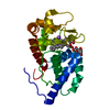
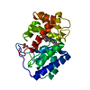
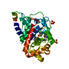
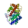
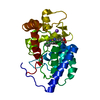
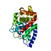
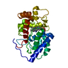
Mass: 28246.775 Da / Num. of mol.: 1 / Fragment: RESIDUES 2-250 / Mutation: YES
Source method: isolated from a genetically manipulated source
Details: HISTIDINE 42 IS IN A DUAL CONFORMATION RESULTING IN A MOVEMENT OF RESIDUES 40-45.
Source: (gene. exp.) GLYCINE MAX (soybean) / Plasmid: SOYAPX/W41A / Production host: ESCHERICHIA COLI (E. coli) / Variant (production host): SG13009 / References: UniProt: Q43758, EC: 1.11.11.1
#2: Chemical ChemComp-HEM / PROTOPORPHYRIN IX CONTAINING FE / HEME / Heme B


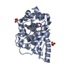
Mass: 616.487 Da / Num. of mol.: 1 / Source method: obtained synthetically / Formula: C34H32FeN4O4
#3: Chemical ChemComp-NA / SODIUM ION


Mass: 22.990 Da / Num. of mol.: 1 / Source method: obtained synthetically / Formula: Na
#4: Water ChemComp-HOH / water / Water


Mass: 18.015 Da / Num. of mol.: 315 / Source method: isolated from a natural source / Formula: H2O
Compound detailsENGINEERED RESIDUE IN CHAIN X, TRP 41 TO ALA

-
Experimental details

-
Experiment

ExperimentMethod: X-RAY DIFFRACTION / Number of used crystals: 1

-
Sample preparation

CrystalDensity Matthews: 1.97 Å3/Da / Density % sol: 37.13 % / Description: NONE
Crystal growpH: 8.3 / Details: pH 8.3

-
Data collection

DiffractionMean temperature: 100 K
Diffraction sourceSource: SYNCHROTRON / Site: ESRF / Beamline: ID14-2 / Wavelength: 0.933
DetectorType: ADSC CCD / Detector: CCD / Date: Mar 3, 2007
RadiationProtocol: SINGLE WAVELENGTH / Monochromatic (M) / Laue (L): M / Scattering type: x-ray
Radiation wavelengthWavelength: 0.933 Å / Relative weight: 1
ReflectionResolution: 1.6→37.8 Å / Num. obs: 34661 / % possible obs: 100 % / Observed criterion σ(I): 3 / Redundancy: 10.18 % / Rmerge(I) obs: 0.09 / Net I/σ(I): 19.5
Reflection shellResolution: 1.6→1.69 Å / Redundancy: 9.89 % / Rmerge(I) obs: 0.66 / Mean I/σ(I) obs: 3.1 / % possible all: 100

-
Processing

Software
NameVersionClassification
REFMAC5.2.0019refinement
MOSFLMdata reduction
SCALAdata scaling
REFMACphasing
RefinementMethod to determine structure: MOLECULAR REPLACEMENT
Starting model: PDB ENTRY 2GGN
Resolution: 1.5→37.8 Å / Cor.coef. Fo:Fc: 0.963 / Cor.coef. Fo:Fc free: 0.952 / SU B: 2.889 / SU ML: 0.049 / Cross valid method: THROUGHOUT / ESU R: 0.093 / ESU R Free: 0.077 / Stereochemistry target values: MAXIMUM LIKELIHOOD / Details: HYDROGENS HAVE BEEN ADDED IN THE RIDING POSITIONS.
RfactorNum. reflection% reflectionSelection details
Rfree0.209 2113 5 %RANDOM
Rwork0.175 ---
obs0.177 39733 100 %-
Solvent computationIon probe radii: 0.8 Å / Shrinkage radii: 0.8 Å / VDW probe radii: 1.2 Å / Solvent model: MASK
Displacement parametersBiso mean: 15.68 Å2
Baniso -1Baniso -2Baniso -3
1--0.13 Å20 Å20 Å2
2---0.13 Å20 Å2
3---0.25 Å2
Refinement stepCycle: LAST / Resolution: 1.5→37.8 Å
ProteinNucleic acidLigandSolventTotal
Num. atoms1874 0 44 315 2233
Refine LS restraints
Refine-IDTypeDev idealDev ideal targetNumber
X-RAY DIFFRACTIONr_bond_refined_d0.010.0222049
X-RAY DIFFRACTIONr_bond_other_d
X-RAY DIFFRACTIONr_angle_refined_deg1.1522.0442804
X-RAY DIFFRACTIONr_angle_other_deg
X-RAY DIFFRACTIONr_dihedral_angle_1_deg4.9765266
X-RAY DIFFRACTIONr_dihedral_angle_2_deg35.67324.09683
X-RAY DIFFRACTIONr_dihedral_angle_3_deg11.23915314
X-RAY DIFFRACTIONr_dihedral_angle_4_deg19.562159
X-RAY DIFFRACTIONr_chiral_restr0.0750.2294
X-RAY DIFFRACTIONr_gen_planes_refined0.0050.021599
X-RAY DIFFRACTIONr_gen_planes_other
X-RAY DIFFRACTIONr_nbd_refined0.2150.21093
X-RAY DIFFRACTIONr_nbd_other
X-RAY DIFFRACTIONr_nbtor_refined0.3050.21398
X-RAY DIFFRACTIONr_nbtor_other
X-RAY DIFFRACTIONr_xyhbond_nbd_refined0.1030.2249
X-RAY DIFFRACTIONr_xyhbond_nbd_other
X-RAY DIFFRACTIONr_metal_ion_refined
X-RAY DIFFRACTIONr_metal_ion_other
X-RAY DIFFRACTIONr_symmetry_vdw_refined0.2130.235
X-RAY DIFFRACTIONr_symmetry_vdw_other
X-RAY DIFFRACTIONr_symmetry_hbond_refined0.1750.225
X-RAY DIFFRACTIONr_symmetry_hbond_other
X-RAY DIFFRACTIONr_symmetry_metal_ion_refined
X-RAY DIFFRACTIONr_symmetry_metal_ion_other
X-RAY DIFFRACTIONr_mcbond_it0.8241.51286
X-RAY DIFFRACTIONr_mcbond_other
X-RAY DIFFRACTIONr_mcangle_it1.34822054
X-RAY DIFFRACTIONr_mcangle_other
X-RAY DIFFRACTIONr_scbond_it1.9583786
X-RAY DIFFRACTIONr_scbond_other
X-RAY DIFFRACTIONr_scangle_it2.9494.5746
X-RAY DIFFRACTIONr_scangle_other
X-RAY DIFFRACTIONr_long_range_B_refined
X-RAY DIFFRACTIONr_long_range_B_other
X-RAY DIFFRACTIONr_rigid_bond_restr
X-RAY DIFFRACTIONr_sphericity_free
X-RAY DIFFRACTIONr_sphericity_bonded
LS refinement shellResolution: 1.5→1.54 Å / Total num. of bins used: 20 /
RfactorNum. reflection
Rfree0.333 163
Rwork0.265 2885

+
About Yorodumi

-
News

-
Feb 9, 2022. New format data for meta-information of EMDB entries

New format data for meta-information of EMDB entries

  • Version 3 of the EMDB header file is now the official format.
  • The previous official version 1.9 will be removed from the archive.

Related info.:EMDB header

External links:wwPDB to switch to version 3 of the EMDB data model

-
Aug 12, 2020. Covid-19 info

Covid-19 info

URL: https://pdbj.org/emnavi/covid19.php

New page: Covid-19 featured information page in EM Navigator.

Related info.:Covid-19 info / Mar 5, 2020. Novel coronavirus structure data

+
Mar 5, 2020. Novel coronavirus structure data

Novel coronavirus structure data

Related info.:Yorodumi Speices / Aug 12, 2020. Covid-19 info

External links:COVID-19 featured content - PDBj / Molecule of the Month (242):Coronavirus Proteases

+
Jan 31, 2019. EMDB accession codes are about to change! (news from PDBe EMDB page)

EMDB accession codes are about to change! (news from PDBe EMDB page)

  • The allocation of 4 digits for EMDB accession codes will soon come to an end. Whilst these codes will remain in use, new EMDB accession codes will include an additional digit and will expand incrementally as the available range of codes is exhausted. The current 4-digit format prefixed with “EMD-” (i.e. EMD-XXXX) will advance to a 5-digit format (i.e. EMD-XXXXX), and so on. It is currently estimated that the 4-digit codes will be depleted around Spring 2019, at which point the 5-digit format will come into force.
  • The EM Navigator/Yorodumi systems omit the EMD- prefix.

Related info.:Q: What is EMD? / ID/Accession-code notation in Yorodumi/EM Navigator

External links:EMDB Accession Codes are Changing Soon! / Contact to PDBj

+
Jul 12, 2017. Major update of PDB

Major update of PDB

  • wwPDB released updated PDB data conforming to the new PDBx/mmCIF dictionary.
  • This is a major update changing the version number from 4 to 5, and with Remediation, in which all the entries are updated.
  • In this update, many items about electron microscopy experimental information are reorganized (e.g. em_software).
  • Now, EM Navigator and Yorodumi are based on the updated data.

External links:wwPDB Remediation / Enriched Model Files Conforming to OneDep Data Standards Now Available in the PDB FTP Archive

-
Yorodumi

Thousand views of thousand structures

  • Yorodumi is a browser for structure data from EMDB, PDB, SASBDB, etc.
  • This page is also the successor to EM Navigator detail page, and also detail information page/front-end page for Omokage search.
  • The word "yorodu" (or yorozu) is an old Japanese word meaning "ten thousand". "mi" (miru) is to see.

Related info.:EMDB / PDB / SASBDB / Comparison of 3 databanks / Yorodumi Search / Aug 31, 2016. New EM Navigator & Yorodumi / Yorodumi Papers / Jmol/JSmol / Function and homology information / Changes in new EM Navigator and Yorodumi

Read more