 Movie
Movie Controller

[English] 日本語
 Yorodumi
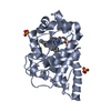
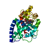
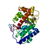
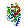
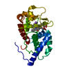
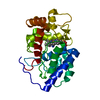
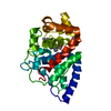
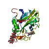
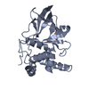
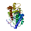
- PDB-1oaf: Ascobate peroxidase from soybean cytosol in complex with ascorbate -
+ Open data

- Basic information
| Entry | Database: PDB / ID: 1oaf | ||||||
|---|---|---|---|---|---|---|---|
| Title | Ascobate peroxidase from soybean cytosol in complex with ascorbate | ||||||
 Components Components | ASCORBATE PEROXIDASE | ||||||
 Keywords Keywords | OXIDOREDUCTASE / HEME PEROXIDASE / PEROXIDE SCAVENGE / ASCORBATE PEROXIDASE | ||||||
| Function / homology |  Function and homology information Function and homology informationL-ascorbate peroxidase / L-ascorbate peroxidase activity / hydrogen peroxide catabolic process / cellular response to oxidative stress / heme binding / metal ion binding Similarity search - Function | ||||||
| Biological species |  | ||||||
| Method |  X-RAY DIFFRACTION / SYNCHROTRON / MOLECULAR REPLACEMENT / Resolution: 1.4 Å X-RAY DIFFRACTION / SYNCHROTRON / MOLECULAR REPLACEMENT / Resolution: 1.4 Å | ||||||
 Authors Authors | Sharp, K.H. / Raven, E.L. / Moody, P.C.E. | ||||||
 Citation Citation | Journal: Nat.Struct.Biol. / Year: 2003 Title: Crystal Structure of the Ascorbate Peroxidase-Ascorbate Complex Authors: Sharp, K.H. / Mewies, M. / Moody, P.C.E. / Raven, E.L. | ||||||
| History |
|
- Structure visualization
| Structure viewer | Molecule: MolmilJmol/JSmol |
|---|
- Downloads & links
-Download
| PDBx/mmCIF format | 1oaf.cif.gz | 131.6 KB | Display | PDBx/mmCIF format |
|---|---|---|---|---|
| PDB format | pdb1oaf.ent.gz | 101.4 KB | Display | PDB format |
| PDBx/mmJSON format | 1oaf.json.gz | Tree view | PDBx/mmJSON format | |
| Others |  Other downloads Other downloads |
-Validation report
| Arichive directory | https://data.pdbj.org/pub/pdb/validation_reports/oa/1oafftp://data.pdbj.org/pub/pdb/validation_reports/oa/1oaf | HTTPS FTP |
|---|
-Related structure data
| Related structure data | 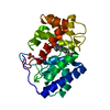 1oagC 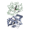 1apxS C: citing same article ( S: Starting model for refinement |
|---|---|
| Similar structure data |
-Links
 PDBj
PDBj
- Assembly
| Deposited unit | 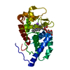
| ||||||||
|---|---|---|---|---|---|---|---|---|---|
| 1 |
| ||||||||
| Unit cell |
|
-Components
| #1: Protein | Mass: 28361.904 Da / Num. of mol.: 1 Source method: isolated from a genetically manipulated source Source: (gene. exp.)  |
|---|---|
| #2: Chemical | ChemComp-HEM /   Mass: 616.487 Da / Num. of mol.: 1 / Source method: obtained synthetically / Formula: C34H32FeN4O4 Mass: 616.487 Da / Num. of mol.: 1 / Source method: obtained synthetically / Formula: C34H32FeN4O4 |
| #3: Chemical | ChemComp-NA /   Mass: 22.990 Da / Num. of mol.: 1 / Source method: obtained synthetically / Formula: Na Mass: 22.990 Da / Num. of mol.: 1 / Source method: obtained synthetically / Formula: Na |
| #4: Sugar | ChemComp-ASC /   Type: L-saccharide / Mass: 176.124 Da / Num. of mol.: 1 / Source method: obtained synthetically / Formula: C6H8O6 Type: L-saccharide / Mass: 176.124 Da / Num. of mol.: 1 / Source method: obtained synthetically / Formula: C6H8O6 |
| #5: Water | ChemComp-HOH /  Mass: 18.015 Da / Num. of mol.: 505 / Source method: isolated from a natural source / Formula: H2O Mass: 18.015 Da / Num. of mol.: 505 / Source method: isolated from a natural source / Formula: H2O |
-Experimental details
-Experiment
| Experiment | Method: X-RAY DIFFRACTION / Number of used crystals: 1 |
|---|
- Sample preparation
| Crystal | Density Matthews: 2.3 Å3/Da / Density % sol: 46 % | ||||||||||||||||||||||||
|---|---|---|---|---|---|---|---|---|---|---|---|---|---|---|---|---|---|---|---|---|---|---|---|---|---|
| Crystal grow | pH: 8.3 / Details: 2.25M LI2SO4, 0.1M HEPES, PH8.3, pH 8.30 | ||||||||||||||||||||||||
| Crystal grow | *PLUS Method: vapor diffusion, sitting drop | ||||||||||||||||||||||||
| Components of the solutions | *PLUS
|
-Data collection
| Diffraction | Mean temperature: 100 K |
|---|---|
| Diffraction source | Source: SYNCHROTRON / Site: ESRF  / Beamline: ID14-4 / Wavelength: 0.94 / Beamline: ID14-4 / Wavelength: 0.94 |
| Detector | Type: ADSC CCD / Detector: CCD / Date: Sep 1, 2002 |
| Radiation | Protocol: SINGLE WAVELENGTH / Monochromatic (M) / Laue (L): M / Scattering type: x-ray |
| Radiation wavelength | Wavelength: 0.94 Å / Relative weight: 1 |
| Reflection | Resolution: 1.4→54 Å / Num. obs: 49370 / % possible obs: 96.2 % / Redundancy: 6.9 % / Rmerge(I) obs: 0.083 / Net I/σ(I): 23.2 |
| Reflection shell | Resolution: 1.4→1.44 Å / Rmerge(I) obs: 0.425 / Mean I/σ(I) obs: 2.9 / % possible all: 96.2 |
| Reflection | *PLUS Highest resolution: 1.4 Å / Lowest resolution: 57.74 Å / Num. measured all: 339792 |
| Reflection shell | *PLUS Highest resolution: 1.4 Å / % possible obs: 96.2 % |
- Processing
| Software |
| ||||||||||||||||||||
|---|---|---|---|---|---|---|---|---|---|---|---|---|---|---|---|---|---|---|---|---|---|
| Refinement | Method to determine structure: MOLECULAR REPLACEMENT Starting model: PDB ENTRY 1APX Resolution: 1.4→57.74 Å / SU B: 0.886 / SU ML: 0.036 / Cross valid method: THROUGHOUT / ESU R: 0.073 / ESU R Free: 0.065 Details: HYDROGENS HAVE BEEN ADDED IN THE RIDING POSITIONS.RESIDUE 1 AND THE N-TERMINAL HIS-TAG ARE NOT VISIBLE
| ||||||||||||||||||||
| Displacement parameters | Biso mean: 15.671 Å2
| ||||||||||||||||||||
| Refinement step | Cycle: LAST / Resolution: 1.4→57.74 Å
| ||||||||||||||||||||
| Refinement | *PLUS Highest resolution: 1.4 Å / Rfactor Rfree: 0.214 / Rfactor Rwork: 0.196 | ||||||||||||||||||||
| Solvent computation | *PLUS | ||||||||||||||||||||
| Displacement parameters | *PLUS | ||||||||||||||||||||
| Refine LS restraints | *PLUS
|
