 Movie
Movie Controller

[English] 日本語
 Yorodumi
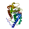
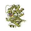
- PDB-1gxy: crystal structure of the eucaryotic mono-ADP-ribosyltransferase A... -
+ Open data

- Basic information
| Entry | Database: PDB / ID: 1gxy | ||||||
|---|---|---|---|---|---|---|---|
| Title | crystal structure of the eucaryotic mono-ADP-ribosyltransferase ART2.2; CRYSTAL FORM A (P21) | ||||||
 Components Components | T-CELL ECTO-ADP-RIBOSYLTRANSFERASE 2 | ||||||
 Keywords Keywords | TRANSFERASE / ADP-RIBOSYLTRANSFERASE / IMMUNO-REGULATION | ||||||
| Function / homology |  Function and homology information Function and homology informationNAD+-protein-arginine ADP-ribosyltransferase / NAD+ glycohydrolase / NAD+-protein-arginine ADP-ribosyltransferase activity / NAD+ nucleosidase activity / NAD+ catabolic process / hydrolase activity, acting on glycosyl bonds / side of membrane / nucleotidyltransferase activity / plasma membrane Similarity search - Function | ||||||
| Biological species |  | ||||||
| Method |  X-RAY DIFFRACTION / SYNCHROTRON / SIRAS / Resolution: 1.71 Å X-RAY DIFFRACTION / SYNCHROTRON / SIRAS / Resolution: 1.71 Å | ||||||
 Authors Authors | Mueller-Dieckmann, C. / Schulz, G.E. | ||||||
 Citation Citation | Journal: J.Mol.Biol. / Year: 2002 Title: Structure of the Ecto-Adp-Ribosyl Transferase Art2.2 From Rat Authors: Mueller-Dieckmann, C. / Ritter, H. / Haag, F. / Koch-Nolte, F. / Schulz, G.E. | ||||||
| History |
|
- Structure visualization
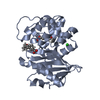
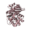
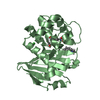
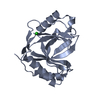
| Structure viewer | Molecule: MolmilJmol/JSmol |
|---|
- Downloads & links
-Download
| PDBx/mmCIF format | 1gxy.cif.gz | 111.8 KB | Display | PDBx/mmCIF format |
|---|---|---|---|---|
| PDB format | pdb1gxy.ent.gz | 86.8 KB | Display | PDB format |
| PDBx/mmJSON format | 1gxy.json.gz | Tree view | PDBx/mmJSON format | |
| Others |  Other downloads Other downloads |
-Validation report
| Arichive directory | https://data.pdbj.org/pub/pdb/validation_reports/gx/1gxyftp://data.pdbj.org/pub/pdb/validation_reports/gx/1gxy | HTTPS FTP |
|---|
-Related structure data


-Links
 PDBj
PDBj- Assembly
| Deposited unit | 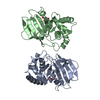
| ||||||||
|---|---|---|---|---|---|---|---|---|---|
| 1 | 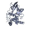
| ||||||||
| 2 | 
| ||||||||
| Unit cell |
| ||||||||
| Noncrystallographic symmetry (NCS) | NCS oper: (Code: given Matrix: (-0.12554, 0.02431, -0.99179), Vector: |
-Components
| #1: Protein | Mass: 26068.447 Da / Num. of mol.: 2 Source method: isolated from a genetically manipulated source Source: (gene. exp.)  References: UniProt: P20974, NAD+-protein-arginine ADP-ribosyltransferase #2: Chemical |   Mass: 92.094 Da / Num. of mol.: 2 / Source method: obtained synthetically / Formula: C3H8O3 Mass: 92.094 Da / Num. of mol.: 2 / Source method: obtained synthetically / Formula: C3H8O3#3: Water | ChemComp-HOH / |  Mass: 18.015 Da / Num. of mol.: 498 / Source method: isolated from a natural source / Formula: H2O Mass: 18.015 Da / Num. of mol.: 498 / Source method: isolated from a natural source / Formula: H2OHas protein modification | Y | |
|---|
-Experimental details
-Experiment
| Experiment | Method: X-RAY DIFFRACTION |
|---|
- Sample preparation
| Crystal | Density Matthews: 2.23 Å3/Da / Density % sol: 48 % | |||||||||||||||||||||||||||||||||||
|---|---|---|---|---|---|---|---|---|---|---|---|---|---|---|---|---|---|---|---|---|---|---|---|---|---|---|---|---|---|---|---|---|---|---|---|---|
| Crystal grow | pH: 8.3 Details: 100 MM TRIS PH8.3, 200 MM LI2SO4, 22 % PEG4000, pH 8.30 | |||||||||||||||||||||||||||||||||||
| Crystal grow | *PLUS Temperature: 293 K / Method: vapor diffusion, hanging dropDetails: Mueller-Dieckmann, C., (2002) Acta Crystallogr., D58, 1211. | |||||||||||||||||||||||||||||||||||
| Components of the solutions | *PLUS
|
-Data collection
| Diffraction | Mean temperature: 100 K |
|---|---|
| Diffraction source | Source: SYNCHROTRON / Site: EMBL/DESY, HAMBURG  / Beamline: X11 / Wavelength: 0.9094 / Beamline: X11 / Wavelength: 0.9094 |
| Radiation | Protocol: SINGLE WAVELENGTH / Monochromatic (M) / Laue (L): M / Scattering type: x-ray |
| Radiation wavelength | Wavelength: 0.9094 Å / Relative weight: 1 |
| Reflection | Resolution: 1.71→29 Å / Num. obs: 48705 / % possible obs: 99.1 % / Redundancy: 4 % / Biso Wilson estimate: 17.5 Å2 / Rsym value: 0.034 / Net I/σ(I): 10.5 |
| Reflection shell | Resolution: 1.71→1.8 Å / Redundancy: 3.8 % / Mean I/σ(I) obs: 6 / Rsym value: 0.119 / % possible all: 99.1 |
| Reflection | *PLUS Highest resolution: 1.7 Å / Lowest resolution: 29 Å / Num. measured all: 195638 / Rmerge(I) obs: 0.034 |
| Reflection shell | *PLUS % possible obs: 99.1 % / Num. unique obs: 6787 / Rmerge(I) obs: 0.119 |
- Processing
| Software | Name: CNS / Classification: refinement | ||||||||||||||||||||||||||||||||||||||||||||||||||||||||||||
|---|---|---|---|---|---|---|---|---|---|---|---|---|---|---|---|---|---|---|---|---|---|---|---|---|---|---|---|---|---|---|---|---|---|---|---|---|---|---|---|---|---|---|---|---|---|---|---|---|---|---|---|---|---|---|---|---|---|---|---|---|---|
| Refinement | Method to determine structure: SIRAS / Resolution: 1.71→29 Å / Rfactor Rfree error: 0.004 / Isotropic thermal model: RESTRAINED / Cross valid method: THROUGHOUT / σ(F): 0 / Details: DISORDERED REGIONS WERE MODELLED STEREOCHEMICALLY
| ||||||||||||||||||||||||||||||||||||||||||||||||||||||||||||
| Solvent computation | Solvent model: FLAT MODEL / Bsol: 50.77 Å2 / ksol: 0.36 e/Å3 | ||||||||||||||||||||||||||||||||||||||||||||||||||||||||||||
| Displacement parameters | Biso mean: 21 Å2
| ||||||||||||||||||||||||||||||||||||||||||||||||||||||||||||
| Refine analyze |
| ||||||||||||||||||||||||||||||||||||||||||||||||||||||||||||
| Refinement step | Cycle: LAST / Resolution: 1.71→29 Å
| ||||||||||||||||||||||||||||||||||||||||||||||||||||||||||||
| Refine LS restraints |
| ||||||||||||||||||||||||||||||||||||||||||||||||||||||||||||
| LS refinement shell | Resolution: 1.71→1.82 Å / Rfactor Rfree error: 0.014 / Total num. of bins used: 6
| ||||||||||||||||||||||||||||||||||||||||||||||||||||||||||||
| Refinement | *PLUS Highest resolution: 1.7 Å / Lowest resolution: 35 Å / % reflection Rfree: 5 % / Rfactor Rfree: 0.223 / Rfactor Rwork: 0.19 | ||||||||||||||||||||||||||||||||||||||||||||||||||||||||||||
| Solvent computation | *PLUS | ||||||||||||||||||||||||||||||||||||||||||||||||||||||||||||
| Displacement parameters | *PLUS Biso mean: 21.1 Å2 | ||||||||||||||||||||||||||||||||||||||||||||||||||||||||||||
| Refine LS restraints | *PLUS
|
