 Movie
Movie Controller

[English] 日本語
 Yorodumi
- PDB-1ecs: THE 1.7 A CRYSTAL STRUCTURE OF A BLEOMYCIN RESISTANCE DETERMINANT... -
+ Open data

- Basic information
| Entry | Database: PDB / ID: 1ecs | ||||||
|---|---|---|---|---|---|---|---|
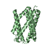
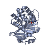
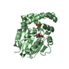
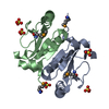
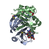
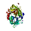
| Title | THE 1.7 A CRYSTAL STRUCTURE OF A BLEOMYCIN RESISTANCE DETERMINANT ENCODED ON THE TRANSPOSON TN5 | ||||||
 Components Components | BLEOMYCIN RESISTANCE PROTEIN | ||||||
 Keywords Keywords | ANTIBIOTIC INHIBITOR / ARM-EXCHANGE | ||||||
| Function / homology |  Function and homology information Function and homology information | ||||||
| Biological species |  Klebsiella pneumoniae (bacteria) Klebsiella pneumoniae (bacteria) | ||||||
| Method |  X-RAY DIFFRACTION / SYNCHROTRON / MOLECULAR REPLACEMENT / Resolution: 1.7 Å X-RAY DIFFRACTION / SYNCHROTRON / MOLECULAR REPLACEMENT / Resolution: 1.7 Å | ||||||
 Authors Authors | Maruyama, M. / Matoba, Y. / Kumagai, T. / Sugiyama, M. | ||||||
 Citation Citation | Journal: J.Biol.Chem. / Year: 2001 Title: Crystal structures of the transposon Tn5-carried bleomycin resistance determinant uncomplexed and complexed with bleomycin. Authors: Maruyama, M. / Kumagai, T. / Matoba, Y. / Hayashida, M. / Fujii, T. / Hata, Y. / Sugiyama, M. #1: Journal: FEBS Lett. / Year: 1999Title: Characterizaton of the bleomycin resistance determinant encoded on the transposon Tn5 Authors: Kumagai, T. / Nakano, T. / Maruyama, M. / Mochizuki, H. / Sugiyama, M. #2: Journal: Acta Crystallogr.,Sect.D / Year: 1999Title: Crystallization and preliminary X-ray diffraction studies of bleomycin-binding protein encoded on the transposon Tn5 Authors: Kumagai, T. / Maruyama, M. / Matoba, Y. / Kawano, Y. / Sugiyama, M. | ||||||
| History |
|
- Structure visualization
| Structure viewer | Molecule: MolmilJmol/JSmol |
|---|
- Downloads & links
-Download
| PDBx/mmCIF format | 1ecs.cif.gz | 63.7 KB | Display | PDBx/mmCIF format |
|---|---|---|---|---|
| PDB format | pdb1ecs.ent.gz | 45.8 KB | Display | PDB format |
| PDBx/mmJSON format | 1ecs.json.gz | Tree view | PDBx/mmJSON format | |
| Others |  Other downloads Other downloads |
-Validation report
| Arichive directory | https://data.pdbj.org/pub/pdb/validation_reports/ec/1ecsftp://data.pdbj.org/pub/pdb/validation_reports/ec/1ecs | HTTPS FTP |
|---|
-Related structure data
| Related structure data |  1ewjC  1qtoS C: citing same article ( S: Starting model for refinement |
|---|---|
| Similar structure data |


-Links
 PDBj
PDBj- Assembly
| Deposited unit | 
| ||||||||||||
|---|---|---|---|---|---|---|---|---|---|---|---|---|---|
| 1 |
| ||||||||||||
| Unit cell |
| ||||||||||||
| Components on special symmetry positions |
|
-Components
| #1: Protein | Mass: 14012.873 Da / Num. of mol.: 2 Source method: isolated from a genetically manipulated source Source: (gene. exp.) Klebsiella pneumoniae (bacteria) / Gene: TRANSPOSON TN5 / Plasmid: PKKTRP / Production host: #2: Chemical | ChemComp-CA / |   Mass: 40.078 Da / Num. of mol.: 1 / Source method: obtained synthetically / Formula: Ca Mass: 40.078 Da / Num. of mol.: 1 / Source method: obtained synthetically / Formula: Ca#3: Chemical |   Mass: 194.226 Da / Num. of mol.: 2 / Source method: obtained synthetically / Formula: C8H18O5 / Comment: precipitant*YM Mass: 194.226 Da / Num. of mol.: 2 / Source method: obtained synthetically / Formula: C8H18O5 / Comment: precipitant*YM#4: Water | ChemComp-HOH / |  Mass: 18.015 Da / Num. of mol.: 111 / Source method: isolated from a natural source / Formula: H2O Mass: 18.015 Da / Num. of mol.: 111 / Source method: isolated from a natural source / Formula: H2O |
|---|
-Experimental details
-Experiment
| Experiment | Method: X-RAY DIFFRACTION / Number of used crystals: 3 |
|---|
- Sample preparation
| Crystal | Density Matthews: 2.42 Å3/Da / Density % sol: 49.2 % | ||||||||||||||||||||
|---|---|---|---|---|---|---|---|---|---|---|---|---|---|---|---|---|---|---|---|---|---|
| Crystal grow | Temperature: 298 K / Method: vapor diffusion, hanging drop / pH: 7 Details: PEG 6000, calcium acetate, sodium cacodylate, pH 7.0, VAPOR DIFFUSION, HANGING DROP, temperature 298.0K | ||||||||||||||||||||
| Crystal | *PLUS Density % sol: 49 % | ||||||||||||||||||||
| Crystal grow | *PLUS | ||||||||||||||||||||
| Components of the solutions | *PLUS
|
-Data collection
| Diffraction | Mean temperature: 298 K |
|---|---|
| Diffraction source | Source: SYNCHROTRON / Site: Photon Factory  / Beamline: BL-18B / Wavelength: 1 / Beamline: BL-18B / Wavelength: 1 |
| Detector | Type: WEISSENBERG / Detector: DIFFRACTOMETER / Date: Feb 20, 1998 |
| Radiation | Protocol: SINGLE WAVELENGTH / Monochromatic (M) / Laue (L): M / Scattering type: x-ray |
| Radiation wavelength | Wavelength: 1 Å / Relative weight: 1 |
| Reflection | Resolution: 1.7→58.77 Å / Num. obs: 28095 / % possible obs: 92.4 % / Observed criterion σ(I): 1 / Redundancy: 10.5 % / Rmerge(I) obs: 0.066 / Net I/σ(I): 6.1 |
| Reflection shell | Resolution: 1.7→1.77 Å / Redundancy: 3.46 % / Rmerge(I) obs: 0.253 / % possible all: 78.1 |
| Reflection | *PLUS Num. measured all: 295760 |
| Reflection shell | *PLUS % possible obs: 78.1 % |
- Processing
| Software |
| |||||||||||||||||||||||||||||||||
|---|---|---|---|---|---|---|---|---|---|---|---|---|---|---|---|---|---|---|---|---|---|---|---|---|---|---|---|---|---|---|---|---|---|---|
| Refinement | Method to determine structure: MOLECULAR REPLACEMENT Starting model: DIMERIC BLMA (1QTO) Resolution: 1.7→5 Å / Num. parameters: 8115 / Num. restraintsaints: 7896 / Stereochemistry target values: ENGH AND HUBER
| |||||||||||||||||||||||||||||||||
| Refine analyze | Occupancy sum hydrogen: 0 / Occupancy sum non hydrogen: 2028.5 | |||||||||||||||||||||||||||||||||
| Refinement step | Cycle: LAST / Resolution: 1.7→5 Å
| |||||||||||||||||||||||||||||||||
| Refine LS restraints |
| |||||||||||||||||||||||||||||||||
| Software | *PLUS Name: SHELXL-97 / Classification: refinement | |||||||||||||||||||||||||||||||||
| Refinement | *PLUS Rfactor obs: 0.192 | |||||||||||||||||||||||||||||||||
| Solvent computation | *PLUS | |||||||||||||||||||||||||||||||||
| Displacement parameters | *PLUS | |||||||||||||||||||||||||||||||||
| Refine LS restraints | *PLUS
|
