 Movie
Movie Controller

[English] 日本語
 Yorodumi
- PDB-1k4k: Crystal structure of E. coli Nicotinic acid mononucleotide adenyl... -
+ Open data

- Basic information
| Entry | Database: PDB / ID: 1k4k | ||||||
|---|---|---|---|---|---|---|---|
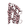
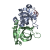
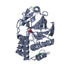
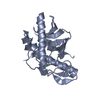
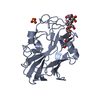
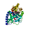
| Title | Crystal structure of E. coli Nicotinic acid mononucleotide adenylyltransferase | ||||||
 Components Components | Nicotinic acid mononucleotide adenylyltransferase | ||||||
 Keywords Keywords | TRANSFERASE / Nucleotidyltransferase | ||||||
| Function / homology |  Function and homology information Function and homology information'de novo' NAD+ biosynthetic process from L-aspartate / nicotinamide-nucleotide adenylyltransferase activity / nicotinate-nucleotide adenylyltransferase / nicotinate-nucleotide adenylyltransferase activity / NAD+ biosynthetic process via the salvage pathway / NAD+ biosynthetic process / ATP binding Similarity search - Function | ||||||
| Biological species |  | ||||||
| Method |  X-RAY DIFFRACTION / SYNCHROTRON / MAD / Resolution: 2 Å X-RAY DIFFRACTION / SYNCHROTRON / MAD / Resolution: 2 Å | ||||||
 Authors Authors | Zhang, H. / Zhou, T. / Kurnasov, O. / Cheek, S. / Grishin, N.V. / Osterman, A.L. | ||||||
 Citation Citation | Journal: Structure / Year: 2002 Title: Crystal structures of E. coli nicotinate mononucleotide adenylyltransferase and its complex with deamido-NAD. Authors: Zhang, H. / Zhou, T. / Kurnasov, O. / Cheek, S. / Grishin, N.V. / Osterman, A. | ||||||
| History |
|
- Structure visualization
| Structure viewer | Molecule: MolmilJmol/JSmol |
|---|
- Downloads & links
-Download
| PDBx/mmCIF format | 1k4k.cif.gz | 188.6 KB | Display | PDBx/mmCIF format |
|---|---|---|---|---|
| PDB format | pdb1k4k.ent.gz | 151.1 KB | Display | PDB format |
| PDBx/mmJSON format | 1k4k.json.gz | Tree view | PDBx/mmJSON format | |
| Others |  Other downloads Other downloads |
-Validation report
| Arichive directory | https://data.pdbj.org/pub/pdb/validation_reports/k4/1k4kftp://data.pdbj.org/pub/pdb/validation_reports/k4/1k4k | HTTPS FTP |
|---|
-Related structure data


-Links
 PDBj
PDBj
- Assembly
| Deposited unit | 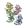
| ||||||||
|---|---|---|---|---|---|---|---|---|---|
| 1 | 
| ||||||||
| 2 | 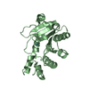
| ||||||||
| 3 | 
| ||||||||
| 4 | 
| ||||||||
| Unit cell |
| ||||||||
| Details | The functional unit of this protein is monomer. |
-Components
| #1: Protein | Mass: 24553.871 Da / Num. of mol.: 4 Source method: isolated from a genetically manipulated source Source: (gene. exp.) References: UniProt: P0A752, nicotinate-nucleotide adenylyltransferase #2: Chemical | ChemComp-XE /   Mass: 131.293 Da / Num. of mol.: 4 / Source method: obtained synthetically / Formula: Xe Mass: 131.293 Da / Num. of mol.: 4 / Source method: obtained synthetically / Formula: Xe#3: Water | ChemComp-HOH / |  Mass: 18.015 Da / Num. of mol.: 585 / Source method: isolated from a natural source / Formula: H2O Mass: 18.015 Da / Num. of mol.: 585 / Source method: isolated from a natural source / Formula: H2O |
|---|
-Experimental details
-Experiment
| Experiment | Method: X-RAY DIFFRACTION / Number of used crystals: 1 |
|---|
- Sample preparation
| Crystal | Density Matthews: 2.57 Å3/Da / Density % sol: 52.07 % | ||||||||||||||||||||||||||||||||||||||||||||||||||||||||
|---|---|---|---|---|---|---|---|---|---|---|---|---|---|---|---|---|---|---|---|---|---|---|---|---|---|---|---|---|---|---|---|---|---|---|---|---|---|---|---|---|---|---|---|---|---|---|---|---|---|---|---|---|---|---|---|---|---|
| Crystal grow | Temperature: 293 K / Method: vapor diffusion, hanging drop / pH: 7 Details: Na Citrate, sodium chloride, Tris, pH 7.0, VAPOR DIFFUSION, HANGING DROP, temperature 20K | ||||||||||||||||||||||||||||||||||||||||||||||||||||||||
| Crystal grow | *PLUS pH: 7.2 | ||||||||||||||||||||||||||||||||||||||||||||||||||||||||
| Components of the solutions | *PLUS
|
-Data collection
| Diffraction |
| ||||||||||||||||||
|---|---|---|---|---|---|---|---|---|---|---|---|---|---|---|---|---|---|---|---|
| Diffraction source |
| ||||||||||||||||||
| Detector |
| ||||||||||||||||||
| Radiation |
| ||||||||||||||||||
| Radiation wavelength |
| ||||||||||||||||||
| Reflection | Resolution: 2→40 Å / Num. all: 66058 / Num. obs: 62463 / % possible obs: 96.4 % / Observed criterion σ(F): 0 / Observed criterion σ(I): -3 / Redundancy: 2.7 % / Rmerge(I) obs: 0.045 / Net I/σ(I): 20.9 | ||||||||||||||||||
| Reflection shell | Resolution: 2→2.03 Å / Rmerge(I) obs: 0.25 / Mean I/σ(I) obs: 4 / % possible all: 81.9 | ||||||||||||||||||
| Reflection | *PLUS Highest resolution: 2 Å / Num. measured all: 169804 / Rmerge(I) obs: 0.045 | ||||||||||||||||||
| Reflection shell | *PLUS % possible obs: 81.9 % / Rmerge(I) obs: 0.25 |

- Processing
| Software |
| ||||||||||||||||||||||||||||||||||||||||||||||||||||||||||||
|---|---|---|---|---|---|---|---|---|---|---|---|---|---|---|---|---|---|---|---|---|---|---|---|---|---|---|---|---|---|---|---|---|---|---|---|---|---|---|---|---|---|---|---|---|---|---|---|---|---|---|---|---|---|---|---|---|---|---|---|---|---|
| Refinement | Method to determine structure: MAD / Resolution: 2→40 Å / Cross valid method: THROUGHOUT / σ(F): 0 / Stereochemistry target values: Engh & Huber
| ||||||||||||||||||||||||||||||||||||||||||||||||||||||||||||
| Refinement step | Cycle: LAST / Resolution: 2→40 Å
| ||||||||||||||||||||||||||||||||||||||||||||||||||||||||||||
| Refine LS restraints |
| ||||||||||||||||||||||||||||||||||||||||||||||||||||||||||||
| LS refinement shell | Highest resolution: 2 Å | ||||||||||||||||||||||||||||||||||||||||||||||||||||||||||||
| Refinement | *PLUS Highest resolution: 2 Å / % reflection Rfree: 5 % / Rfactor obs: 0.21 / Rfactor Rfree: 0.262 / Rfactor Rwork: 0.21 | ||||||||||||||||||||||||||||||||||||||||||||||||||||||||||||
| Solvent computation | *PLUS | ||||||||||||||||||||||||||||||||||||||||||||||||||||||||||||
| Displacement parameters | *PLUS | ||||||||||||||||||||||||||||||||||||||||||||||||||||||||||||
| Refine LS restraints | *PLUS Type: o_angle_deg / Dev ideal: 1.7 |
