 Movie
Movie Controller


+ Open data

- Basic information
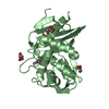
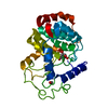
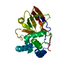
| Entry | Database: PDB / ID: 6ydc | ||||||
|---|---|---|---|---|---|---|---|
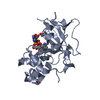
| Title | X-ray structure of LPMO | ||||||
 Components Components | LPMO lytic polysaccharide monooxygenase | ||||||
 Keywords Keywords | METAL BINDING PROTEIN / Lytic Polysaccharide Monooxygenase / Complex / cellotetraose | ||||||
| Function / homology |  Function and homology information Function and homology informationlytic cellulose monooxygenase (C4-dehydrogenating) / cellulose catabolic process / monooxygenase activity / extracellular region / metal ion binding Similarity search - Function | ||||||
| Biological species |  Collariella virescens (fungus) Collariella virescens (fungus) | ||||||
| Method |  X-RAY DIFFRACTION / SYNCHROTRON / MOLECULAR REPLACEMENT / Resolution: 2 Å X-RAY DIFFRACTION / SYNCHROTRON / MOLECULAR REPLACEMENT / Resolution: 2 Å | ||||||
 Authors Authors | Tandrup, T. / Tryfona, T. / Frandsen, K.E.H. / Johansen, K.S. / Dupree, P. / Lo Leggio, L. | ||||||
| Funding support |  Denmark, 1items Denmark, 1items
| ||||||
 Citation Citation | Journal: Biochemistry / Year: 2020 Title: Oligosaccharide Binding and Thermostability of Two Related AA9 Lytic Polysaccharide Monooxygenases. Authors: Tandrup, T. / Tryfona, T. / Frandsen, K.E.H. / Johansen, K.S. / Dupree, P. / Lo Leggio, L. | ||||||
| History |
|
- Structure visualization
| Structure viewer | Molecule: MolmilJmol/JSmol |
|---|
- Downloads & links
-Download
| PDBx/mmCIF format | 6ydc.cif.gz | 226.8 KB | Display | PDBx/mmCIF format |
|---|---|---|---|---|
| PDB format | pdb6ydc.ent.gz | 179.5 KB | Display | PDB format |
| PDBx/mmJSON format | 6ydc.json.gz | Tree view | PDBx/mmJSON format | |
| Others |  Other downloads Other downloads |
-Validation report
| Arichive directory | https://data.pdbj.org/pub/pdb/validation_reports/yd/6ydcftp://data.pdbj.org/pub/pdb/validation_reports/yd/6ydc | HTTPS FTP |
|---|
-Related structure data
| Related structure data |  6yddC  6ydeC  6ydfC  6ydgC  5achS S: Starting model for refinement C: citing same article ( |
|---|---|
| Similar structure data |

-Links
 PDBj
PDBj- Assembly
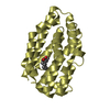
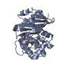
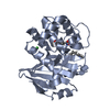
| Deposited unit | 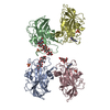
| ||||||||
|---|---|---|---|---|---|---|---|---|---|
| 1 | 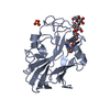
| ||||||||
| 2 | 
| ||||||||
| 3 | 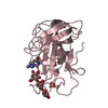
| ||||||||
| 4 | 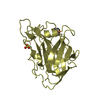
| ||||||||
| Unit cell |
|
-Components
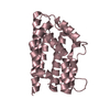
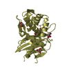
-Protein , 1 types, 4 molecules ABCD
| #1: Protein | Mass: 27791.133 Da / Num. of mol.: 4 Source method: isolated from a genetically manipulated source Source: (gene. exp.) Collariella virescens (fungus) / Gene: aa9 / Production host: |
|---|
-Sugars , 2 types, 3 molecules
| #2: Polysaccharide | 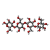  Source method: isolated from a genetically manipulated source Details: oligosaccharide / References: beta-cellotetraose #3: Polysaccharide | beta-D-glucopyranose-(1-4)-beta-D-glucopyranose-(1-4)-beta-D-glucopyranose / beta-cellotriose |   Source method: isolated from a genetically manipulated source Details: oligosaccharide / References: beta-cellotriose |
|---|
-Non-polymers , 3 types, 1464 molecules 


| #4: Chemical | ChemComp-CU /  Mass: 63.546 Da / Num. of mol.: 4 / Source method: obtained synthetically / Formula: Cu / Feature type: SUBJECT OF INVESTIGATION Mass: 63.546 Da / Num. of mol.: 4 / Source method: obtained synthetically / Formula: Cu / Feature type: SUBJECT OF INVESTIGATION#5: Chemical | ChemComp-SO4 /  Mass: 96.063 Da / Num. of mol.: 4 / Source method: obtained synthetically / Formula: SO4 Mass: 96.063 Da / Num. of mol.: 4 / Source method: obtained synthetically / Formula: SO4#6: Water | ChemComp-HOH / | Mass: 18.015 Da / Num. of mol.: 1456 / Source method: isolated from a natural source / Formula: H2O |
|---|
-Details
| Has ligand of interest | Y |
|---|
-Experimental details
-Experiment
| Experiment | Method: X-RAY DIFFRACTION / Number of used crystals: 1 |
|---|
- Sample preparation
| Crystal | Density Matthews: 2.95 Å3/Da / Density % sol: 58.25 % |
|---|---|
| Crystal grow | Temperature: 298.15 K / Method: vapor diffusion, sitting drop / pH: 6.5 / Details: 0.1 M NaCl, 0.1 M HEPES pH 6.5, 1.5 M (NH4)2SO4 |
-Data collection
| Diffraction | Mean temperature: 100 K / Serial crystal experiment: N |
|---|---|
| Diffraction source | Source: SYNCHROTRON / Site: ESRF  / Beamline: MASSIF-3 / Wavelength: 0.96 Å / Beamline: MASSIF-3 / Wavelength: 0.96 Å |
| Detector | Type: DECTRIS EIGER X 4M / Detector: PIXEL / Date: Feb 18, 2017 |
| Radiation | Protocol: SINGLE WAVELENGTH / Monochromatic (M) / Laue (L): M / Scattering type: x-ray |
| Radiation wavelength | Wavelength: 0.96 Å / Relative weight: 1 |
| Reflection | Resolution: 2→50 Å / Num. obs: 85419 / % possible obs: 98.4 % / Redundancy: 4.55 % / CC1/2: 0.989 / Net I/σ(I): 5.54 |
| Reflection shell | Resolution: 2→2.05 Å / Redundancy: 4.16 % / Mean I/σ(I) obs: 1.44 / Num. unique obs: 6184 / CC1/2: 0.544 / % possible all: 97.2 |
- Processing
| Software |
| ||||||||||||||||||
|---|---|---|---|---|---|---|---|---|---|---|---|---|---|---|---|---|---|---|---|
| Refinement | Method to determine structure: MOLECULAR REPLACEMENT Starting model: 5ACH Resolution: 2→50 Å / Cross valid method: FREE R-VALUE
| ||||||||||||||||||
| Displacement parameters | Biso max: 61.24 Å2 / Biso mean: 14.8197 Å2 / Biso min: 2.27 Å2 | ||||||||||||||||||
| Refinement step | Cycle: LAST / Resolution: 2→50 Å
|
